 Movie
Movie Controller

[English] 日本語
 Yorodumi
- PDB-1tjb: Crystal Structure of a High Affinity Lanthanide-Binding Peptide (LBT) -
+ Open data

- Basic information
| Entry | Database: PDB / ID: 1tjb | ||||||
|---|---|---|---|---|---|---|---|
| Title | Crystal Structure of a High Affinity Lanthanide-Binding Peptide (LBT) | ||||||
 Components Components | Lanthanide-Binding Peptide | ||||||
 Keywords Keywords | DE NOVO PROTEIN / Lanthanide-Based Resonance Energy Transfer / Fluorescence / EF-Hand / Troponin Based Design / Lanthanide Binding Tag | ||||||
| Function / homology | TERBIUM(III) ION Function and homology information Function and homology information | ||||||
| Method |  X-RAY DIFFRACTION / SAD PHASING / Resolution: 2 Å X-RAY DIFFRACTION / SAD PHASING / Resolution: 2 Å | ||||||
 Authors Authors | Nitz, M. / Sherawat, M. / Franz, K.J. / Peisach, E. / Allen, K.N. / Imperiali, B. | ||||||
 Citation Citation | Journal: Angew.Chem.Int.Ed.Engl. / Year: 2004 Title: Structural Origin of the High Affinity of a Chemically Evolved Lanthanide-Binding Peptide Authors: Nitz, M. / Sherawat, M. / Franz, K.J. / Peisach, E. / Allen, K.N. / Imperiali, B. #1: Journal: Chembiochem / Year: 2003Title: Lanthanide-binding tags as versatile protein coexpression probes Authors: Franz, K.J. / Nitz, M. / Imperiali, B. | ||||||
| History |
|
- Structure visualization
| Structure viewer | Molecule: MolmilJmol/JSmol |
|---|
- Downloads & links
-Download
| PDBx/mmCIF format | 1tjb.cif.gz | 17.8 KB | Display | PDBx/mmCIF format |
|---|---|---|---|---|
| PDB format | pdb1tjb.ent.gz | 11 KB | Display | PDB format |
| PDBx/mmJSON format | 1tjb.json.gz | Tree view | PDBx/mmJSON format | |
| Others |  Other downloads Other downloads |
-Validation report
| Arichive directory | https://data.pdbj.org/pub/pdb/validation_reports/tj/1tjbftp://data.pdbj.org/pub/pdb/validation_reports/tj/1tjb | HTTPS FTP |
|---|
-Related structure data
| Related structure data | |
|---|---|
| Similar structure data |

-Links
 PDBj
PDBj
- Assembly
| Deposited unit | 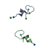
| ||||||||
|---|---|---|---|---|---|---|---|---|---|
| 1 | 
| ||||||||
| 2 | 
| ||||||||
| Unit cell |
| ||||||||
| Details | The solution assembly is a monomer |
-Components
| #1: Protein/peptide | Mass: 1986.036 Da / Num. of mol.: 2 / Source method: obtained synthetically Details: Peptide prepared by automated solid phase peptide synthesis #2: Chemical |   Mass: 158.925 Da / Num. of mol.: 2 / Source method: obtained synthetically / Formula: Tb Mass: 158.925 Da / Num. of mol.: 2 / Source method: obtained synthetically / Formula: Tb#3: Chemical |   Mass: 35.453 Da / Num. of mol.: 3 / Source method: obtained synthetically / Formula: Cl Mass: 35.453 Da / Num. of mol.: 3 / Source method: obtained synthetically / Formula: Cl#4: Water | ChemComp-HOH / |  Mass: 18.015 Da / Num. of mol.: 45 / Source method: isolated from a natural source / Formula: H2O Mass: 18.015 Da / Num. of mol.: 45 / Source method: isolated from a natural source / Formula: H2OHas protein modification | Y | |
|---|
-Experimental details
-Experiment
| Experiment | Method: X-RAY DIFFRACTION / Number of used crystals: 1 |
|---|
- Sample preparation
| Crystal | Density Matthews: 2.3 Å3/Da / Density % sol: 46 % |
|---|---|
| Crystal grow | Temperature: 298 K / Method: vapor diffusion, sitting drop / pH: 6.5 Details: t-Butanol, Terbium Chloride, sodium chloride, tris, pH 6.5, VAPOR DIFFUSION, SITTING DROP, temperature 298K |
-Data collection
| Diffraction | Mean temperature: 100 K |
|---|---|
| Diffraction source | Source: ROTATING ANODE / Type: RIGAKU RU300 / Wavelength: 1.5418 Å |
| Detector | Type: RIGAKU RAXIS IV / Detector: IMAGE PLATE / Date: Jul 26, 2003 / Details: Osmic Mirrors |
| Radiation | Monochromator: OSMIC MIRRORS / Protocol: SINGLE WAVELENGTH / Monochromatic (M) / Laue (L): M / Scattering type: x-ray |
| Radiation wavelength | Wavelength: 1.5418 Å / Relative weight: 1 |
| Reflection | Resolution: 2→55 Å / Num. all: 2315 / Num. obs: 2315 / % possible obs: 99.1 % / Observed criterion σ(F): 0 / Observed criterion σ(I): 0 / Redundancy: 3.6 % / Biso Wilson estimate: 6.8 Å2 / Rmerge(I) obs: 0.107 / Net I/σ(I): 15.2 |
| Reflection shell | Resolution: 2→2.07 Å / Redundancy: 3.8 % / Rmerge(I) obs: 0.171 / Mean I/σ(I) obs: 7.8 / Num. unique all: 212 / % possible all: 100 |
- Processing
| Software |
| |||||||||||||||||||||||||||||||||||||||||||||||||||||||||||||||
|---|---|---|---|---|---|---|---|---|---|---|---|---|---|---|---|---|---|---|---|---|---|---|---|---|---|---|---|---|---|---|---|---|---|---|---|---|---|---|---|---|---|---|---|---|---|---|---|---|---|---|---|---|---|---|---|---|---|---|---|---|---|---|---|---|
| Refinement | Method to determine structure: SAD PHASING / Resolution: 2→17.18 Å / Isotropic thermal model: isotropic / Cross valid method: THROUGHOUT / σ(F): 0 / Stereochemistry target values: Engh & Huber
| |||||||||||||||||||||||||||||||||||||||||||||||||||||||||||||||
| Displacement parameters | Biso mean: 14.77 Å2 | |||||||||||||||||||||||||||||||||||||||||||||||||||||||||||||||
| Refine analyze |
| |||||||||||||||||||||||||||||||||||||||||||||||||||||||||||||||
| Refinement step | Cycle: LAST / Resolution: 2→17.18 Å
| |||||||||||||||||||||||||||||||||||||||||||||||||||||||||||||||
| Refine LS restraints |
| |||||||||||||||||||||||||||||||||||||||||||||||||||||||||||||||
| LS refinement shell | Refine-ID: X-RAY DIFFRACTION
| |||||||||||||||||||||||||||||||||||||||||||||||||||||||||||||||
| Xplor file |
|
