 Movie
Movie Controller


+ Open data

- Basic information
| Entry | Database: PDB / ID: 1t68 | ||||||
|---|---|---|---|---|---|---|---|
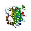
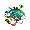
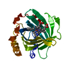
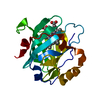
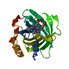
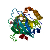
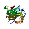
| Title | Crystal structure of nitrophorin 2 complex with NO | ||||||
 Components Components | Nitrophorin 2 | ||||||
 Keywords Keywords | TRANSPORT PROTEIN / beta barrel / lipocalin / heme / nitric oxide / ruffling | ||||||
| Function / homology |  Function and homology information Function and homology informationhistamine binding / nitric oxide binding / vasodilation / toxin activity / extracellular region / metal ion binding Similarity search - Function | ||||||
| Biological species |  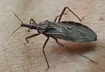 Rhodnius prolixus (insect) Rhodnius prolixus (insect) | ||||||
| Method |  X-RAY DIFFRACTION / SYNCHROTRON / FOURIER SYNTHESIS / Resolution: 1.45 Å X-RAY DIFFRACTION / SYNCHROTRON / FOURIER SYNTHESIS / Resolution: 1.45 Å | ||||||
 Authors Authors | Weichsel, A. / Montfort, W.R. | ||||||
 Citation Citation | Journal: To be Published Title: Conformational change and heme ruffling in nitrophorin 2, a nitric oxide carrier from kissing bug Authors: Weichsel, A. / Montfort, W.R. #1: Journal: J.Biol.Chem. / Year: 2000Title: The crystal structure of nitrophorin 2. A trifunctional antihemostatic protein from the saliva of Rhodnius prolixus. Authors: Andersen, J.F. / Montfort, W.R. #2: Journal: Biochemistry / Year: 2001Title: Ligand-induced heme ruffling and bent NO geometry in ultra-high-resolution structures of nitrophorin 4 Authors: Roberts, S.A. / Weichsel, A. / Qui, Y. / Shelnutt, J.A. / Walker, F.A. / Montfort, W.R. | ||||||
| History |
|
- Structure visualization
| Structure viewer | Molecule: MolmilJmol/JSmol |
|---|
- Downloads & links
-Download
| PDBx/mmCIF format | 1t68.cif.gz | 89.8 KB | Display | PDBx/mmCIF format |
|---|---|---|---|---|
| PDB format | pdb1t68.ent.gz | 67.8 KB | Display | PDB format |
| PDBx/mmJSON format | 1t68.json.gz | Tree view | PDBx/mmJSON format | |
| Others |  Other downloads Other downloads |
-Validation report
| Arichive directory | https://data.pdbj.org/pub/pdb/validation_reports/t6/1t68ftp://data.pdbj.org/pub/pdb/validation_reports/t6/1t68 | HTTPS FTP |
|---|
-Related structure data
| Related structure data |  1pm1C  1euoS S: Starting model for refinement C: citing same article ( |
|---|---|
| Similar structure data |


-Links
 PDBj
PDBj


- Assembly
| Deposited unit | 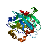
| ||||||||||||
|---|---|---|---|---|---|---|---|---|---|---|---|---|---|
| 1 |
| ||||||||||||
| Unit cell |
| ||||||||||||
| Components on special symmetry positions |
|

-Components
| #1: Protein | Mass: 20080.391 Da / Num. of mol.: 1 Source method: isolated from a genetically manipulated source Source: (gene. exp.) Rhodnius prolixus (insect) / Plasmid: pET17B / Species (production host): Escherichia coli / Production host:  |
|---|---|
| #2: Chemical | ChemComp-HEM /   Mass: 616.487 Da / Num. of mol.: 1 / Source method: obtained synthetically / Formula: C34H32FeN4O4 Mass: 616.487 Da / Num. of mol.: 1 / Source method: obtained synthetically / Formula: C34H32FeN4O4 |
| #3: Chemical | ChemComp-NO /   Mass: 30.006 Da / Num. of mol.: 1 / Source method: obtained synthetically / Formula: NO Mass: 30.006 Da / Num. of mol.: 1 / Source method: obtained synthetically / Formula: NO |
| #4: Water | ChemComp-HOH /  Mass: 18.015 Da / Num. of mol.: 102 / Source method: isolated from a natural source / Formula: H2O Mass: 18.015 Da / Num. of mol.: 102 / Source method: isolated from a natural source / Formula: H2O |
| Has protein modification | Y |
-Experimental details
-Experiment
| Experiment | Method: X-RAY DIFFRACTION / Number of used crystals: 1 |
|---|
- Sample preparation
| Crystal | Density Matthews: 2 Å3/Da / Density % sol: 39.4 % |
|---|---|
| Crystal grow | Temperature: 298 K / Method: vapor diffusion, hanging drop / pH: 7.5 Details: sodium citrate, pH 7.5, VAPOR DIFFUSION, HANGING DROP, temperature 298K |
-Data collection
| Diffraction | Mean temperature: 100 K |
|---|---|
| Diffraction source | Source: SYNCHROTRON / Site: APS  / Beamline: 14-BM-C / Wavelength: 0.9 Å / Beamline: 14-BM-C / Wavelength: 0.9 Å |
| Detector | Type: ADSC QUANTUM 4 / Detector: CCD / Date: Apr 15, 2003 / Details: bent Si-mirror |
| Radiation | Monochromator: bent Ge(111) / Protocol: SINGLE WAVELENGTH / Monochromatic (M) / Laue (L): M / Scattering type: x-ray |
| Radiation wavelength | Wavelength: 0.9 Å / Relative weight: 1 |
| Reflection | Resolution: 1.45→22.8 Å / Num. all: 26809 / Num. obs: 26809 / % possible obs: 89.3 % / Observed criterion σ(F): 0 / Observed criterion σ(I): 0 / Redundancy: 4.4 % / Biso Wilson estimate: 17.1 Å2 / Rmerge(I) obs: 0.093 / Net I/σ(I): 13.2 |
| Reflection shell | Resolution: 1.45→1.5 Å / Redundancy: 4.5 % / Rmerge(I) obs: 0.246 / Mean I/σ(I) obs: 2.3 / Num. unique all: 2879 / % possible all: 98.1 |
- Processing
| Software |
| ||||||||||||||||||||||||||||||||||||||||||||||||||||||||||||||||||||||||||||||||||||||||||||||||||||||||||||||||||||||||
|---|---|---|---|---|---|---|---|---|---|---|---|---|---|---|---|---|---|---|---|---|---|---|---|---|---|---|---|---|---|---|---|---|---|---|---|---|---|---|---|---|---|---|---|---|---|---|---|---|---|---|---|---|---|---|---|---|---|---|---|---|---|---|---|---|---|---|---|---|---|---|---|---|---|---|---|---|---|---|---|---|---|---|---|---|---|---|---|---|---|---|---|---|---|---|---|---|---|---|---|---|---|---|---|---|---|---|---|---|---|---|---|---|---|---|---|---|---|---|---|---|---|
| Refinement | Method to determine structure: FOURIER SYNTHESIS Starting model: PDB ENTRY 1EUO Resolution: 1.45→22.8 Å / Cor.coef. Fo:Fc: 0.943 / Cor.coef. Fo:Fc free: 0.929 / SU B: 1.284 / SU ML: 0.051 / Isotropic thermal model: anisotropic / Cross valid method: THROUGHOUT / σ(F): 0 / σ(I): 0 / ESU R: 0.109 / ESU R Free: 0.089 / Stereochemistry target values: MAXIMUM LIKELIHOOD / Details: HYDROGENS HAVE BEEN ADDED IN THE RIDING POSITIONS
| ||||||||||||||||||||||||||||||||||||||||||||||||||||||||||||||||||||||||||||||||||||||||||||||||||||||||||||||||||||||||
| Solvent computation | Ion probe radii: 0.8 Å / Shrinkage radii: 0.8 Å / VDW probe radii: 1.4 Å / Solvent model: BABINET MODEL WITH MASK | ||||||||||||||||||||||||||||||||||||||||||||||||||||||||||||||||||||||||||||||||||||||||||||||||||||||||||||||||||||||||
| Displacement parameters | Biso mean: 20.273 Å2
| ||||||||||||||||||||||||||||||||||||||||||||||||||||||||||||||||||||||||||||||||||||||||||||||||||||||||||||||||||||||||
| Refine analyze |
| ||||||||||||||||||||||||||||||||||||||||||||||||||||||||||||||||||||||||||||||||||||||||||||||||||||||||||||||||||||||||
| Refinement step | Cycle: LAST / Resolution: 1.45→22.8 Å
| ||||||||||||||||||||||||||||||||||||||||||||||||||||||||||||||||||||||||||||||||||||||||||||||||||||||||||||||||||||||||
| Refine LS restraints |
| ||||||||||||||||||||||||||||||||||||||||||||||||||||||||||||||||||||||||||||||||||||||||||||||||||||||||||||||||||||||||
| LS refinement shell | Resolution: 1.45→1.488 Å / Total num. of bins used: 20
|
