- PDB-1t3u: Unknown conserved bacterial protein from Pseudomonas aeruginosa PAO1 -
+
Open data
ID or keywords:
Loading...
-
Basic information
Entry
Database: PDB / ID: 1t3u
Title
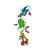
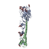
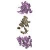
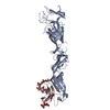
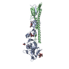
Unknown conserved bacterial protein from Pseudomonas aeruginosa PAO1
Components
conserved hypothetical protein
Keywords
STRUCTURAL GENOMICS / UNKNOWN FUNCTION / T1445 / NYSGXRC / UNKNOWN ORF / COG3027 / PSI / Protein Structure Initiative / New York SGX Research Center for Structural Genomics
Function / homology
Function and homology information
septin ring assembly / cell septum / division septum assembly / FtsZ-dependent cytokinesis / cell division site / cytosol Similarity search - Function
Cell division protein ZapA protomer, N-terminal domain / Cell division protein ZapA, N-terminal / Cell division protein ZapA-like / Cell division protein ZapA-like superfamily / Cell division protein ZapA / Single alpha-helices involved in coiled-coils or other helix-helix interfaces - #50 / Double Stranded RNA Binding Domain / Single alpha-helices involved in coiled-coils or other helix-helix interfaces / Up-down Bundle / 2-Layer Sandwich ...Cell division protein ZapA protomer, N-terminal domain / Cell division protein ZapA, N-terminal / Cell division protein ZapA-like / Cell division protein ZapA-like superfamily / Cell division protein ZapA / Single alpha-helices involved in coiled-coils or other helix-helix interfaces - #50 / Double Stranded RNA Binding Domain / Single alpha-helices involved in coiled-coils or other helix-helix interfaces / Up-down Bundle / 2-Layer Sandwich / Mainly Alpha / Alpha Beta Similarity search - Domain/homology
THIS ENTRY CONTAINS THE CRYSTALLOGRAPHIC ASYMMETRIC UNIT WHICH CONSISTS OF 4 CHAIN(S). THE ...THIS ENTRY CONTAINS THE CRYSTALLOGRAPHIC ASYMMETRIC UNIT WHICH CONSISTS OF 4 CHAIN(S). THE BIOLOGICAL MOLECULE IS REPRESENTED AS A TETRAMER IN THE ASYMMETRIC UNIT, HOWEVER THE PHYSIOLOGICAL STATE OF THE PROTEIN IS NOT KNOWN YET.
In the structure databanks used in Yorodumi, some data are registered as the other names, "COVID-19 virus" and "2019-nCoV". Here are the details of the virus and the list of structure data.
Jan 31, 2019. EMDB accession codes are about to change! (news from PDBe EMDB page)
EMDB accession codes are about to change! (news from PDBe EMDB page)
The allocation of 4 digits for EMDB accession codes will soon come to an end. Whilst these codes will remain in use, new EMDB accession codes will include an additional digit and will expand incrementally as the available range of codes is exhausted. The current 4-digit format prefixed with “EMD-” (i.e. EMD-XXXX) will advance to a 5-digit format (i.e. EMD-XXXXX), and so on. It is currently estimated that the 4-digit codes will be depleted around Spring 2019, at which point the 5-digit format will come into force.
The EM Navigator/Yorodumi systems omit the EMD- prefix.
Related info.:Q: What is EMD? / ID/Accession-code notation in Yorodumi/EM Navigator
Yorodumi is a browser for structure data from EMDB, PDB, SASBDB, etc.
This page is also the successor to EM Navigator detail page, and also detail information page/front-end page for Omokage search.
The word "yorodu" (or yorozu) is an old Japanese word meaning "ten thousand". "mi" (miru) is to see.
Related info.:EMDB / PDB / SASBDB / Comparison of 3 databanks / Yorodumi Search / Aug 31, 2016. New EM Navigator & Yorodumi / Yorodumi Papers / Jmol/JSmol / Function and homology information / Changes in new EM Navigator and Yorodumi
 Movie
Movie Controller

 Yorodumi
 Open data
 Basic information
 Components
 Keywords
 Function and homology information
 Pseudomonas aeruginosa PAO1 (bacteria)
 X-RAY DIFFRACTION /
  Authors
 Citation
 Structure visualization
 Downloads & links
 Other downloads
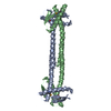


 PDBj
PDBj Assembly
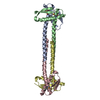
 Mass: 18.015 Da / Num. of mol.: 48 / Source method: isolated from a natural source / Formula: H2O
 Sample preparation
 / Beamline: 31-ID / Wavelength: 0.979 Å
 Processing