 Movie
Movie Controller

[English] 日本語
 Yorodumi
- PDB-1t0d: Crystal Structure of 2-aminopurine labelled bacterial decoding si... -
+ Open data

- Basic information
| Entry | Database: PDB / ID: 1t0d | ||||||
|---|---|---|---|---|---|---|---|
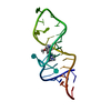
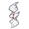
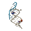
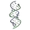
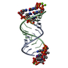
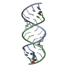
| Title | Crystal Structure of 2-aminopurine labelled bacterial decoding site RNA | ||||||
 Components Components |
| ||||||
 Keywords Keywords | RNA / 2-AMINOPURINE / BACTERIAL DECODING SITE RNA / FLUORESCENCE EMISSION SPECTRA / 9-beta-D-Ribofuranosyl-9H-purin-2-amine | ||||||
| Function / homology | RNA / RNA (> 10) Function and homology information Function and homology information | ||||||
| Method |  X-RAY DIFFRACTION / MOLECULAR REPLACEMENT / Resolution: 2.2 Å X-RAY DIFFRACTION / MOLECULAR REPLACEMENT / Resolution: 2.2 Å | ||||||
 Authors Authors | Shandrick, S. / Zhao, Q. / Han, Q. / Ayida, B.K. / Takahashi, M. / Winters, G.C. / Simonsen, K.B. / Vourloumis, D. / Hermann, T. | ||||||
 Citation Citation | Journal: Angew.Chem.Int.Ed.Engl. / Year: 2004 Title: Monitoring molecular recognition of the ribosomal decoding site. Authors: Shandrick, S. / Zhao, Q. / Han, Q. / Ayida, B.K. / Takahashi, M. / Winters, G.C. / Simonsen, K.B. / Vourloumis, D. / Hermann, T. | ||||||
| History |
|
- Structure visualization
| Structure viewer | Molecule: MolmilJmol/JSmol |
|---|
- Downloads & links
-Download
| PDBx/mmCIF format | 1t0d.cif.gz | 49.8 KB | Display | PDBx/mmCIF format |
|---|---|---|---|---|
| PDB format | pdb1t0d.ent.gz | 35.6 KB | Display | PDB format |
| PDBx/mmJSON format | 1t0d.json.gz | Tree view | PDBx/mmJSON format | |
| Others |  Other downloads Other downloads |
-Validation report
| Arichive directory | https://data.pdbj.org/pub/pdb/validation_reports/t0/1t0dftp://data.pdbj.org/pub/pdb/validation_reports/t0/1t0d | HTTPS FTP |
|---|
-Related structure data


-Links
 PDBj
PDBj


- Assembly
| Deposited unit | 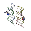
| ||||||||
|---|---|---|---|---|---|---|---|---|---|
| 1 | 
| ||||||||
| 2 | 
| ||||||||
| Unit cell |
|
-Components
| #1: RNA chain | Mass: 5555.333 Da / Num. of mol.: 2 / Source method: obtained synthetically #2: RNA chain | Mass: 5015.079 Da / Num. of mol.: 2 / Source method: obtained synthetically #3: Water | ChemComp-HOH / |  Mass: 18.015 Da / Num. of mol.: 215 / Source method: isolated from a natural source / Formula: H2O Mass: 18.015 Da / Num. of mol.: 215 / Source method: isolated from a natural source / Formula: H2O |
|---|
-Experimental details
-Experiment
| Experiment | Method: X-RAY DIFFRACTION / Number of used crystals: 1 |
|---|
- Sample preparation
| Crystal | Density Matthews: 2.1 Å3/Da / Density % sol: 40.5 % |
|---|---|
| Crystal grow | Temperature: 293.15 K / Method: vapor diffusion, hanging drop / pH: 6.8 Details: Ammonium sulfate, magnesium acetate, cacodylate, pH 6.8, VAPOR DIFFUSION, HANGING DROP, temperature 293.15K |
-Data collection
| Diffraction | Mean temperature: 103 K |
|---|---|
| Diffraction source | Source: ROTATING ANODE / Type: RIGAKU / Wavelength: 1.5418 Å |
| Detector | Type: RIGAKU RAXIS IV / Detector: IMAGE PLATE / Date: Jun 14, 2003 / Details: mirrors |
| Radiation | Monochromator: Ni FILTER / Protocol: SINGLE WAVELENGTH / Monochromatic (M) / Laue (L): M / Scattering type: x-ray |
| Radiation wavelength | Wavelength: 1.5418 Å / Relative weight: 1 |
| Reflection | Resolution: 2.2→30 Å / Num. all: 8984 / Num. obs: 8903 / % possible obs: 99.1 % / Observed criterion σ(F): 3 / Observed criterion σ(I): 3 / Redundancy: 8.6 % / Biso Wilson estimate: 19.7 Å2 / Rmerge(I) obs: 0.09 / Net I/σ(I): 15.3 |
| Reflection shell | Resolution: 2.2→2.28 Å / Rmerge(I) obs: 0.311 / Mean I/σ(I) obs: 3.3 / Num. unique all: 844 / % possible all: 96.1 |
- Processing
| Software |
| |||||||||||||||||||||||||
|---|---|---|---|---|---|---|---|---|---|---|---|---|---|---|---|---|---|---|---|---|---|---|---|---|---|---|
| Refinement | Method to determine structure: MOLECULAR REPLACEMENT Starting model: a model of RNA Resolution: 2.2→8 Å / Isotropic thermal model: anisotropic / Cross valid method: THROUGHOUT / σ(F): 0 / σ(I): 0 / Stereochemistry target values: Engh & Huber
| |||||||||||||||||||||||||
| Displacement parameters | Biso mean: 25.8 Å2
| |||||||||||||||||||||||||
| Refine analyze |
| |||||||||||||||||||||||||
| Refinement step | Cycle: LAST / Resolution: 2.2→8 Å
| |||||||||||||||||||||||||
| Refine LS restraints |
| |||||||||||||||||||||||||
| LS refinement shell | Resolution: 2.2→2.34 Å / Rfactor Rfree error: 0.031
|
