 Movie
Movie Controller

[English] 日本語
 Yorodumi
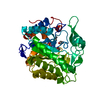
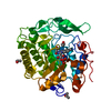
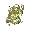
- PDB-1so0: Crystal structure of human galactose mutarotase complexed with ga... -
+ Open data

- Basic information
| Entry | Database: PDB / ID: 1so0 | ||||||
|---|---|---|---|---|---|---|---|
| Title | Crystal structure of human galactose mutarotase complexed with galactose | ||||||
 Components Components | aldose 1-epimerase | ||||||
 Keywords Keywords | ISOMERASE / mutartoase / epimerase / galactosemia | ||||||
| Function / homology |  Function and homology information Function and homology informationDefective GALM causes GALAC4 / aldose 1-epimerase / aldose 1-epimerase activity / galactose metabolic process / beta-D-galactose catabolic process via UDP-galactose, Leloir pathway / Galactose catabolism / glucose metabolic process / carbohydrate binding / carbohydrate metabolic process / extracellular exosome / cytosol Similarity search - Function | ||||||
| Biological species |  Homo sapiens (human) Homo sapiens (human) | ||||||
| Method |  X-RAY DIFFRACTION / MOLECULAR REPLACEMENT / Resolution: 2.3 Å X-RAY DIFFRACTION / MOLECULAR REPLACEMENT / Resolution: 2.3 Å | ||||||
 Authors Authors | Thoden, J.B. / Timson, D.J. / Reece, R.J. / Holden, H.M. | ||||||
 Citation Citation | Journal: J.Biol.Chem. / Year: 2004 Title: Molecular structure of human galactose mutarotase Authors: Thoden, J.B. / Timson, D.J. / Reece, R.J. / Holden, H.M. | ||||||
| History |
|
- Structure visualization
| Structure viewer | Molecule: MolmilJmol/JSmol |
|---|
- Downloads & links
-Download
| PDBx/mmCIF format | 1so0.cif.gz | 281.8 KB | Display | PDBx/mmCIF format |
|---|---|---|---|---|
| PDB format | pdb1so0.ent.gz | 226.7 KB | Display | PDB format |
| PDBx/mmJSON format | 1so0.json.gz | Tree view | PDBx/mmJSON format | |
| Others |  Other downloads Other downloads |
-Validation report
| Arichive directory | https://data.pdbj.org/pub/pdb/validation_reports/so/1so0ftp://data.pdbj.org/pub/pdb/validation_reports/so/1so0 | HTTPS FTP |
|---|
-Related structure data
| Related structure data |  1snzSC S: Starting model for refinement C: citing same article ( |
|---|---|
| Similar structure data |


-Links
 PDBj
PDBj


- Assembly
| Deposited unit | 
| ||||||||
|---|---|---|---|---|---|---|---|---|---|
| 1 | 
| ||||||||
| 2 | 
| ||||||||
| 3 | 
| ||||||||
| 4 | 
| ||||||||
| Unit cell |
|
-Components
| #1: Protein | Mass: 38005.602 Da / Num. of mol.: 4 Source method: isolated from a genetically manipulated source Source: (gene. exp.) Homo sapiens (human) / Gene: GALM / Plasmid: pTYB12 / Production host:  #2: Sugar | ChemComp-GAL /   Type: D-saccharide, beta linking / Mass: 180.156 Da / Num. of mol.: 4 Type: D-saccharide, beta linking / Mass: 180.156 Da / Num. of mol.: 4Source method: isolated from a genetically manipulated source Formula: C6H12O6 #3: Water | ChemComp-HOH / |  Mass: 18.015 Da / Num. of mol.: 406 / Source method: isolated from a natural source / Formula: H2O Mass: 18.015 Da / Num. of mol.: 406 / Source method: isolated from a natural source / Formula: H2O |
|---|
-Experimental details
-Experiment
| Experiment | Method: X-RAY DIFFRACTION / Number of used crystals: 1 |
|---|
- Sample preparation
| Crystal | Density Matthews: 2.47 Å3/Da / Density % sol: 50.1 % |
|---|---|
| Crystal grow | Temperature: 275 K / Method: batch / pH: 6 Details: PEG 8000, sodium chloride, lithium chloride, galactose, MES, pH 6.0, batch, temperature 275K |
-Data collection
| Diffraction | Mean temperature: 275 K |
|---|---|
| Diffraction source | Source: ROTATING ANODE / Type: RIGAKU RU200 / Wavelength: 1.5418 Å |
| Detector | Type: SIEMENS HI-STAR / Detector: AREA DETECTOR / Date: Feb 10, 2004 / Details: super long mirrors |
| Radiation | Monochromator: Ni FILTER / Protocol: SINGLE WAVELENGTH / Monochromatic (M) / Laue (L): M / Scattering type: x-ray |
| Radiation wavelength | Wavelength: 1.5418 Å / Relative weight: 1 |
| Reflection | Resolution: 2.3→30 Å / Num. all: 61156 / Num. obs: 61156 / % possible obs: 94.7 % / Observed criterion σ(F): 0 / Observed criterion σ(I): 0 / Redundancy: 2.1 % / Rsym value: 0.07 / Net I/σ(I): 10.8 |
| Reflection shell | Resolution: 2.3→2.4 Å / Redundancy: 1.4 % / Mean I/σ(I) obs: 2.8 / Num. unique all: 5979 / Rsym value: 0.264 / % possible all: 77.5 |
- Processing
| Software |
| |||||||||||||||||||||||||
|---|---|---|---|---|---|---|---|---|---|---|---|---|---|---|---|---|---|---|---|---|---|---|---|---|---|---|
| Refinement | Method to determine structure: MOLECULAR REPLACEMENT Starting model: PDB entry 1snz Resolution: 2.3→30 Å / Cross valid method: THROUGHOUT / σ(F): 0 / Stereochemistry target values: Engh & Huber
| |||||||||||||||||||||||||
| Refinement step | Cycle: LAST / Resolution: 2.3→30 Å
| |||||||||||||||||||||||||
| Refine LS restraints |
|
