| 登録情報 | データベース: PDB / ID: 1sk4
|
|---|
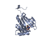
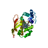
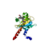
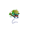
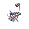
| タイトル | crystal structure of the C-terminal peptidoglycan-binding domain of human peptidoglycan recognition protein Ialpha |
|---|
 要素 要素 | Peptidoglycan recognition protein I-alpha |
|---|
 キーワード キーワード | IMMUNE SYSTEM / alpha/beta mix |
|---|
| 機能・相同性 |  機能・相同性情報 機能・相同性情報
negative regulation of natural killer cell differentiation involved in immune response / peptidoglycan immune receptor activity / N-acetylmuramoyl-L-alanine amidase activity / peptidoglycan binding / biological process involved in interaction with host / Antimicrobial peptides / negative regulation of type II interferon production / detection of bacterium / peptidoglycan catabolic process / antimicrobial humoral immune response mediated by antimicrobial peptide ...negative regulation of natural killer cell differentiation involved in immune response / peptidoglycan immune receptor activity / N-acetylmuramoyl-L-alanine amidase activity / peptidoglycan binding / biological process involved in interaction with host / Antimicrobial peptides / negative regulation of type II interferon production / detection of bacterium / peptidoglycan catabolic process / antimicrobial humoral immune response mediated by antimicrobial peptide / killing of cells of another organism / defense response to Gram-positive bacterium / defense response to bacterium / immune response / protein heterodimerization activity / innate immune response / protein-containing complex / extracellular region / zinc ion binding / membrane類似検索 - 分子機能 Peptidoglycan recognition protein family domain, metazoa/bacteria / Peptidoglycan recognition protein / Animal peptidoglycan recognition proteins homologous to Bacteriophage T3 lysozyme. / Lysozyme-like / Peptidoglycan recognition protein-like / Ami_2 / N-acetylmuramoyl-L-alanine amidase / N-acetylmuramoyl-L-alanine amidase domain / N-acetylmuramoyl-L-alanine amidase/PGRP domain superfamily / 3-Layer(aba) Sandwich / Alpha Beta類似検索 - ドメイン・相同性 |
|---|
| 生物種 |  Homo sapiens (ヒト) Homo sapiens (ヒト) |
|---|
| 手法 |  X線回折 / シンクロトロン / 分子置換 / 解像度: 1.65 Å X線回折 / シンクロトロン / 分子置換 / 解像度: 1.65 Å |
|---|
 データ登録者 データ登録者 | Guan, R. / Malchiodi, E.L. / Qian, W. / Schuck, P. / Mariuzza, R.A. |
|---|
 引用 引用 | ジャーナル: J.Biol.Chem. / 年: 2004
タイトル: Crystal structure of the C-terminal peptidoglycan-binding domain of human peptidoglycan recognition protein Ialpha
著者: Guan, R. / Malchiodi, E.L. / Qian, W. / Schuck, P. / Mariuzza, R.A. |
|---|
| 履歴 | | 登録 | 2004年3月4日 | 登録サイト: RCSB / 処理サイト: RCSB |
|---|
| 改定 1.0 | 2004年7月13日 | Provider: repository / タイプ: Initial release |
|---|
| 改定 1.1 | 2008年4月29日 | Group: Version format compliance |
|---|
| 改定 1.2 | 2011年7月13日 | Group: Derived calculations / Version format compliance |
|---|
| 改定 1.3 | 2023年8月23日 | Group: Data collection / Database references ...Data collection / Database references / Derived calculations / Refinement description
カテゴリ: chem_comp_atom / chem_comp_bond ...chem_comp_atom / chem_comp_bond / database_2 / pdbx_initial_refinement_model / pdbx_struct_conn_angle / struct_conn / struct_ref_seq_dif / struct_site
Item: _database_2.pdbx_DOI / _database_2.pdbx_database_accession ..._database_2.pdbx_DOI / _database_2.pdbx_database_accession / _pdbx_struct_conn_angle.ptnr1_auth_comp_id / _pdbx_struct_conn_angle.ptnr1_auth_seq_id / _pdbx_struct_conn_angle.ptnr1_label_asym_id / _pdbx_struct_conn_angle.ptnr1_label_atom_id / _pdbx_struct_conn_angle.ptnr1_label_comp_id / _pdbx_struct_conn_angle.ptnr1_label_seq_id / _pdbx_struct_conn_angle.ptnr3_auth_comp_id / _pdbx_struct_conn_angle.ptnr3_auth_seq_id / _pdbx_struct_conn_angle.ptnr3_label_asym_id / _pdbx_struct_conn_angle.ptnr3_label_atom_id / _pdbx_struct_conn_angle.ptnr3_label_comp_id / _pdbx_struct_conn_angle.ptnr3_label_seq_id / _pdbx_struct_conn_angle.value / _struct_conn.pdbx_dist_value / _struct_conn.pdbx_leaving_atom_flag / _struct_conn.ptnr1_auth_comp_id / _struct_conn.ptnr1_auth_seq_id / _struct_conn.ptnr1_label_asym_id / _struct_conn.ptnr1_label_atom_id / _struct_conn.ptnr1_label_comp_id / _struct_conn.ptnr1_label_seq_id / _struct_conn.ptnr2_auth_comp_id / _struct_conn.ptnr2_auth_seq_id / _struct_conn.ptnr2_label_asym_id / _struct_conn.ptnr2_label_atom_id / _struct_conn.ptnr2_label_comp_id / _struct_conn.ptnr2_label_seq_id / _struct_ref_seq_dif.details / _struct_site.pdbx_auth_asym_id / _struct_site.pdbx_auth_comp_id / _struct_site.pdbx_auth_seq_id |
|---|
| 改定 1.4 | 2023年11月15日 | Group: Data collection / カテゴリ: chem_comp_atom / chem_comp_bond / Item: _chem_comp_atom.atom_id / _chem_comp_bond.atom_id_2 |
|---|
| 改定 1.5 | 2024年11月13日 | Group: Structure summary
カテゴリ: pdbx_entry_details / pdbx_modification_feature |
|---|
|
|---|
 ムービー
ムービー コントローラー


 データを開く
 基本情報
基本情報 構造の表示
構造の表示 ダウンロードとリンク
 その他のダウンロード


 PDBj
PDBj

 集合体
集合体


 分子量: 22.990 Da / 分子数: 1 / 由来タイプ: 合成 / 式: Na
 分子量: 18.015 Da / 分子数: 137 / 由来タイプ: 天然 / 式: H2O
 試料調製
試料調製 / ビームライン: X26C / 波長: 1 Å
 解析
解析