[English] 日本語
Yorodumi
- PDB-1sk4: crystal structure of the C-terminal peptidoglycan-binding domain ... -

+
Open data


ID or keywords:

Loading...

-
Basic information

Entry
Database: PDB / ID: 1sk4
Title
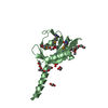
crystal structure of the C-terminal peptidoglycan-binding domain of human peptidoglycan recognition protein Ialpha
ComponentsPeptidoglycan recognition protein I-alpha
KeywordsIMMUNE SYSTEM / alpha/beta mix
Function / homology
Function and homology information


negative regulation of natural killer cell differentiation involved in immune response / peptidoglycan immune receptor activity / N-acetylmuramoyl-L-alanine amidase activity / biological process involved in interaction with host / peptidoglycan binding / Antimicrobial peptides / negative regulation of type II interferon production / detection of bacterium / peptidoglycan catabolic process / antimicrobial humoral immune response mediated by antimicrobial peptide ...negative regulation of natural killer cell differentiation involved in immune response / peptidoglycan immune receptor activity / N-acetylmuramoyl-L-alanine amidase activity / biological process involved in interaction with host / peptidoglycan binding / Antimicrobial peptides / negative regulation of type II interferon production / detection of bacterium / peptidoglycan catabolic process / antimicrobial humoral immune response mediated by antimicrobial peptide / killing of cells of another organism / defense response to Gram-positive bacterium / protein heterodimerization activity / innate immune response / protein-containing complex / zinc ion binding / extracellular region / membrane
Similarity search - Function
Peptidoglycan recognition protein family domain, metazoa/bacteria / Peptidoglycan recognition protein / Animal peptidoglycan recognition proteins homologous to Bacteriophage T3 lysozyme. / Lysozyme-like / Peptidoglycan recognition protein-like / Ami_2 / N-acetylmuramoyl-L-alanine amidase / N-acetylmuramoyl-L-alanine amidase domain / N-acetylmuramoyl-L-alanine amidase/PGRP domain superfamily / 3-Layer(aba) Sandwich / Alpha Beta
Similarity search - Domain/homology
Peptidoglycan recognition protein 3
Similarity search - Component
Biological speciesHomo sapiens (human)
MethodX-RAY DIFFRACTION / SYNCHROTRON / MOLECULAR REPLACEMENT / Resolution: 1.65 Å
AuthorsGuan, R. / Malchiodi, E.L. / Qian, W. / Schuck, P. / Mariuzza, R.A.
CitationJournal: J.Biol.Chem. / Year: 2004
Title: Crystal structure of the C-terminal peptidoglycan-binding domain of human peptidoglycan recognition protein Ialpha
Authors: Guan, R. / Malchiodi, E.L. / Qian, W. / Schuck, P. / Mariuzza, R.A.
History
DepositionMar 4, 2004Deposition site: RCSB / Processing site: RCSB
Revision 1.0Jul 13, 2004Provider: repository / Type: Initial release
Revision 1.1Apr 29, 2008Group: Version format compliance
Revision 1.2Jul 13, 2011Group: Derived calculations / Version format compliance
Revision 1.3Aug 23, 2023Group: Data collection / Database references ...Data collection / Database references / Derived calculations / Refinement description
Category: chem_comp_atom / chem_comp_bond ...chem_comp_atom / chem_comp_bond / database_2 / pdbx_initial_refinement_model / pdbx_struct_conn_angle / struct_conn / struct_ref_seq_dif / struct_site
Item: _database_2.pdbx_DOI / _database_2.pdbx_database_accession ..._database_2.pdbx_DOI / _database_2.pdbx_database_accession / _pdbx_struct_conn_angle.ptnr1_auth_comp_id / _pdbx_struct_conn_angle.ptnr1_auth_seq_id / _pdbx_struct_conn_angle.ptnr1_label_asym_id / _pdbx_struct_conn_angle.ptnr1_label_atom_id / _pdbx_struct_conn_angle.ptnr1_label_comp_id / _pdbx_struct_conn_angle.ptnr1_label_seq_id / _pdbx_struct_conn_angle.ptnr3_auth_comp_id / _pdbx_struct_conn_angle.ptnr3_auth_seq_id / _pdbx_struct_conn_angle.ptnr3_label_asym_id / _pdbx_struct_conn_angle.ptnr3_label_atom_id / _pdbx_struct_conn_angle.ptnr3_label_comp_id / _pdbx_struct_conn_angle.ptnr3_label_seq_id / _pdbx_struct_conn_angle.value / _struct_conn.pdbx_dist_value / _struct_conn.pdbx_leaving_atom_flag / _struct_conn.ptnr1_auth_comp_id / _struct_conn.ptnr1_auth_seq_id / _struct_conn.ptnr1_label_asym_id / _struct_conn.ptnr1_label_atom_id / _struct_conn.ptnr1_label_comp_id / _struct_conn.ptnr1_label_seq_id / _struct_conn.ptnr2_auth_comp_id / _struct_conn.ptnr2_auth_seq_id / _struct_conn.ptnr2_label_asym_id / _struct_conn.ptnr2_label_atom_id / _struct_conn.ptnr2_label_comp_id / _struct_conn.ptnr2_label_seq_id / _struct_ref_seq_dif.details / _struct_site.pdbx_auth_asym_id / _struct_site.pdbx_auth_comp_id / _struct_site.pdbx_auth_seq_id
Revision 1.4Nov 15, 2023Group: Data collection / Category: chem_comp_atom / chem_comp_bond / Item: _chem_comp_atom.atom_id / _chem_comp_bond.atom_id_2

-
Structure visualization

Structure viewerMolecule:
MolmilJmol/JSmol

Downloads & links

-
Assembly

Deposited unit
A: Peptidoglycan recognition protein I-alpha
hetero molecules


Theoretical massNumber of molelcules
Total (without water)18,1092
Polymers18,0861
Non-polymers231
Water2,468137
1


  • Idetical with deposited unit
  • defined by author&software
TypeNameSymmetry operationNumber
identity operation1_555x,y,z1
MethodPQS
2
A: Peptidoglycan recognition protein I-alpha
hetero molecules

A: Peptidoglycan recognition protein I-alpha
hetero molecules


Theoretical massNumber of molelcules
Total (without water)36,2174
Polymers36,1712
Non-polymers462
Water362
TypeNameSymmetry operationNumber
identity operation1_555x,y,z1
crystal symmetry operation6_765-x+2,-x+y+1,-z+2/31
Buried area4220 Å2
ΔGint-56 kcal/mol
Surface area14180 Å2
MethodPISA
Unit cell
Length a, b, c (Å)64.464, 64.464, 63.996
Angle α, β, γ (deg.)90.00, 90.00, 120.00
Int Tables number154
Space group name H-MP3221
Components on special symmetry positions
IDModelComponents
11A-55-

HOH

-
Components

#1: Protein Peptidoglycan recognition protein I-alpha / Peptidoglycan recognition protein intermediate alpha / PGRP-I-alpha / PGLYRPIalpha


Mass: 18085.520 Da / Num. of mol.: 1
Source method: isolated from a genetically manipulated source
Source: (gene. exp.) Homo sapiens (human) / Gene: PGRPIA / Plasmid: pT7-7 / Species (production host): Escherichia coli / Production host: Escherichia coli BL21(DE3) (bacteria) / Strain (production host): BL21 DE3 / References: UniProt: Q96LB9
#2: Chemical ChemComp-NA / SODIUM ION


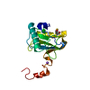
Mass: 22.990 Da / Num. of mol.: 1 / Source method: obtained synthetically / Formula: Na
#3: Water ChemComp-HOH / water


Mass: 18.015 Da / Num. of mol.: 137 / Source method: isolated from a natural source / Formula: H2O

-
Experimental details

-
Experiment

ExperimentMethod: X-RAY DIFFRACTION / Number of used crystals: 1

-
Sample preparation

CrystalDensity Matthews: 2.15 Å3/Da / Density % sol: 42.35 %
Crystal growTemperature: 298 K / Method: vapor diffusion, hanging drop / pH: 7
Details: sodium potassium tartrate, Zinc chloride, Tritox x-100, pH 7.0, VAPOR DIFFUSION, HANGING DROP, temperature 298K

-
Data collection

DiffractionMean temperature: 100 K
Diffraction sourceSource: SYNCHROTRON / Site: NSLS / Beamline: X26C / Wavelength: 1 Å
DetectorType: ADSC QUANTUM 4 / Detector: CCD / Date: Nov 18, 2003
RadiationMonochromator: Si / Protocol: SINGLE WAVELENGTH / Monochromatic (M) / Laue (L): M / Scattering type: x-ray
Radiation wavelengthWavelength: 1 Å / Relative weight: 1
ReflectionResolution: 1.65→30 Å / Num. all: 18925 / Num. obs: 18925 / % possible obs: 99.9 % / Observed criterion σ(F): 0 / Observed criterion σ(I): 0 / Biso Wilson estimate: 19.5 Å2 / Rmerge(I) obs: 0.066
Reflection shellResolution: 1.65→1.71 Å / Rmerge(I) obs: 0.5 / Num. unique all: 1837 / % possible all: 99.8

-
Processing

Software
NameVersionClassification
CNS1.1refinement
HKL-2000data reduction
SCALEPACKdata scaling
AMoREphasing
RefinementMethod to determine structure: MOLECULAR REPLACEMENT
Starting model: 1OHT
Resolution: 1.65→28.79 Å / Rfactor Rfree error: 0.008 / Data cutoff high absF: 1237895.3 / Data cutoff low absF: 0 / Isotropic thermal model: RESTRAINED / Cross valid method: THROUGHOUT / σ(F): 0 / Stereochemistry target values: Engh & Huber
RfactorNum. reflection% reflectionSelection details
Rfree0.212 753 4 %RANDOM
Rwork0.196 ---
all0.196 18900 --
obs0.196 18900 99.8 %-
Solvent computationSolvent model: FLAT MODEL / Bsol: 40.9213 Å2 / ksol: 0.357945 e/Å3
Displacement parametersBiso mean: 21.8 Å2
Baniso -1Baniso -2Baniso -3
1--2.46 Å2-0.84 Å20 Å2
2---2.46 Å20 Å2
3---4.92 Å2
Refine analyze
FreeObs
Luzzati coordinate error0.22 Å0.19 Å
Luzzati d res low-5 Å
Luzzati sigma a0.14 Å0.13 Å
Refinement stepCycle: LAST / Resolution: 1.65→28.79 Å
ProteinNucleic acidLigandSolventTotal
Num. atoms1274 0 1 137 1412
Refine LS restraints
Refine-IDTypeDev ideal
X-RAY DIFFRACTIONc_bond_d0.006
X-RAY DIFFRACTIONc_angle_deg1.4
X-RAY DIFFRACTIONc_dihedral_angle_d23.2
X-RAY DIFFRACTIONc_improper_angle_d0.81
LS refinement shellResolution: 1.65→1.75 Å / Rfactor Rfree error: 0.023 / Total num. of bins used: 6
RfactorNum. reflection% reflection
Rfree0.265 138 4.5 %
Rwork0.236 2934 -
obs--99.8 %
Xplor file
Refine-IDSerial noParam fileTopol file
X-RAY DIFFRACTION1PROTEIN_OCYS_REP.PARAMPROTEIN_OCYS.TOP
X-RAY DIFFRACTION2ION.PARAM
X-RAY DIFFRACTION3WATER_REP.PARAM

+
About Yorodumi

-
News

-
Feb 9, 2022. New format data for meta-information of EMDB entries

New format data for meta-information of EMDB entries

  • Version 3 of the EMDB header file is now the official format.
  • The previous official version 1.9 will be removed from the archive.

Related info.:EMDB header

External links:wwPDB to switch to version 3 of the EMDB data model

-
Aug 12, 2020. Covid-19 info

Covid-19 info

URL: https://pdbjlvh1.pdbj.org/emnavi/covid19.php

New page: Covid-19 featured information page in EM Navigator.

Related info.:Covid-19 info / Mar 5, 2020. Novel coronavirus structure data

+
Mar 5, 2020. Novel coronavirus structure data

Novel coronavirus structure data

Related info.:Yorodumi Speices / Aug 12, 2020. Covid-19 info

External links:COVID-19 featured content - PDBj / Molecule of the Month (242):Coronavirus Proteases

+
Jan 31, 2019. EMDB accession codes are about to change! (news from PDBe EMDB page)

EMDB accession codes are about to change! (news from PDBe EMDB page)

  • The allocation of 4 digits for EMDB accession codes will soon come to an end. Whilst these codes will remain in use, new EMDB accession codes will include an additional digit and will expand incrementally as the available range of codes is exhausted. The current 4-digit format prefixed with “EMD-” (i.e. EMD-XXXX) will advance to a 5-digit format (i.e. EMD-XXXXX), and so on. It is currently estimated that the 4-digit codes will be depleted around Spring 2019, at which point the 5-digit format will come into force.
  • The EM Navigator/Yorodumi systems omit the EMD- prefix.

Related info.:Q: What is EMD? / ID/Accession-code notation in Yorodumi/EM Navigator

External links:EMDB Accession Codes are Changing Soon! / Contact to PDBj

+
Jul 12, 2017. Major update of PDB

Major update of PDB

  • wwPDB released updated PDB data conforming to the new PDBx/mmCIF dictionary.
  • This is a major update changing the version number from 4 to 5, and with Remediation, in which all the entries are updated.
  • In this update, many items about electron microscopy experimental information are reorganized (e.g. em_software).
  • Now, EM Navigator and Yorodumi are based on the updated data.

External links:wwPDB Remediation / Enriched Model Files Conforming to OneDep Data Standards Now Available in the PDB FTP Archive

-
Yorodumi

Thousand views of thousand structures

  • Yorodumi is a browser for structure data from EMDB, PDB, SASBDB, etc.
  • This page is also the successor to EM Navigator detail page, and also detail information page/front-end page for Omokage search.
  • The word "yorodu" (or yorozu) is an old Japanese word meaning "ten thousand". "mi" (miru) is to see.

Related info.:EMDB / PDB / SASBDB / Comparison of 3 databanks / Yorodumi Search / Aug 31, 2016. New EM Navigator & Yorodumi / Yorodumi Papers / Jmol/JSmol / Function and homology information / Changes in new EM Navigator and Yorodumi

Read more