 Movie
Movie Controller

[English] 日本語
 Yorodumi
- PDB-1sjd: x-ray structure of o-succinylbenzoate synthase complexed with n-s... -
+ Open data

- Basic information
| Entry | Database: PDB / ID: 1sjd | ||||||
|---|---|---|---|---|---|---|---|
| Title | x-ray structure of o-succinylbenzoate synthase complexed with n-succinyl phenylglycine | ||||||
 Components Components | N-acylamino acid racemase | ||||||
 Keywords Keywords | LYASE / ISOMERASE / racemase | ||||||
| Function / homology |  Function and homology information Function and homology informationo-succinylbenzoate synthase / O-succinylbenzoate synthase activity / Isomerases; Racemases and epimerases; Acting on amino acids and derivatives / racemase and epimerase activity / menaquinone biosynthetic process / metal ion binding Similarity search - Function | ||||||
| Biological species |  Amycolatopsis sp. (bacteria) Amycolatopsis sp. (bacteria) | ||||||
| Method |  X-RAY DIFFRACTION / SYNCHROTRON / FOURIER SYNTHESIS / Resolution: 1.87 Å X-RAY DIFFRACTION / SYNCHROTRON / FOURIER SYNTHESIS / Resolution: 1.87 Å | ||||||
 Authors Authors | Thoden, J.B. / Taylor-Ringia, E.A. / Garrett, J.B. / Gerlt, J.A. / Holden, H.M. / Rayment, I. | ||||||
 Citation Citation | Journal: Biochemistry / Year: 2004 Title: Evolution of Enzymatic Activity in the Enolase Superfamily: Structural Studies of the Promiscuous o-Succinylbenzoate Synthase from Amycolatopsis Authors: Thoden, J.B. / Taylor-Ringia, E.A. / Garrett, J.B. / Gerlt, J.A. / Holden, H.M. / Rayment, I. | ||||||
| History |
|
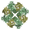
- Structure visualization
| Structure viewer | Molecule: MolmilJmol/JSmol |
|---|
- Downloads & links
-Download
| PDBx/mmCIF format | 1sjd.cif.gz | 311.2 KB | Display | PDBx/mmCIF format |
|---|---|---|---|---|
| PDB format | pdb1sjd.ent.gz | 250.5 KB | Display | PDB format |
| PDBx/mmJSON format | 1sjd.json.gz | Tree view | PDBx/mmJSON format | |
| Others |  Other downloads Other downloads |
-Validation report
| Arichive directory | https://data.pdbj.org/pub/pdb/validation_reports/sj/1sjdftp://data.pdbj.org/pub/pdb/validation_reports/sj/1sjd | HTTPS FTP |
|---|
-Related structure data
| Related structure data | 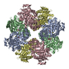 1sjaC  1sjbSC  1sjcC C: citing same article ( S: Starting model for refinement |
|---|---|
| Similar structure data |
-Links
 PDBj
PDBj
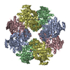
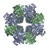
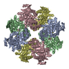
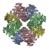
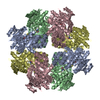
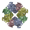
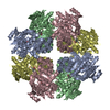
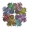
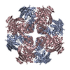
- Assembly
| Deposited unit | 
| ||||||||
|---|---|---|---|---|---|---|---|---|---|
| 1 | 
| ||||||||
| Unit cell |
|
-Components
| #1: Protein | Mass: 39448.270 Da / Num. of mol.: 4 Source method: isolated from a genetically manipulated source Source: (gene. exp.) Amycolatopsis sp. (bacteria) / Gene: AAAR / Plasmid: pET17b / Species (production host): Escherichia coli / Production host: #2: Chemical | ChemComp-NPG /   Mass: 251.235 Da / Num. of mol.: 6 / Source method: obtained synthetically / Formula: C12H13NO5 Mass: 251.235 Da / Num. of mol.: 6 / Source method: obtained synthetically / Formula: C12H13NO5#3: Water | ChemComp-HOH / |  Mass: 18.015 Da / Num. of mol.: 1162 / Source method: isolated from a natural source / Formula: H2O Mass: 18.015 Da / Num. of mol.: 1162 / Source method: isolated from a natural source / Formula: H2O |
|---|
-Experimental details
-Experiment
| Experiment | Method: X-RAY DIFFRACTION / Number of used crystals: 1 |
|---|
- Sample preparation
| Crystal | Density Matthews: 3.64 Å3/Da / Density % sol: 66.16 % |
|---|---|
| Crystal grow | Temperature: 298 K / Method: batch / pH: 8 Details: PEG 8000, HEPES, MgCl2, n-succinylphenylglycine, pH 8.0, batch, temperature 298K |
-Data collection
| Diffraction | Mean temperature: 100 K |
|---|---|
| Diffraction source | Source: SYNCHROTRON / Site: APS  / Beamline: 19-ID / Wavelength: 0.6525 Å / Beamline: 19-ID / Wavelength: 0.6525 Å |
| Detector | Detector: CCD / Date: Apr 7, 2001 |
| Radiation | Protocol: SINGLE WAVELENGTH / Monochromatic (M) / Laue (L): M / Scattering type: x-ray |
| Radiation wavelength | Wavelength: 0.6525 Å / Relative weight: 1 |
| Reflection | Resolution: 1.87→50 Å / Num. all: 186822 / Num. obs: 186822 / % possible obs: 99.7 % / Observed criterion σ(F): 0 / Observed criterion σ(I): 0 / Redundancy: 10.1 % / Rsym value: 0.085 / Net I/σ(I): 40.1 |
| Reflection shell | Resolution: 1.87→1.94 Å / Redundancy: 5.2 % / Mean I/σ(I) obs: 3.3 / Rsym value: 0.377 / % possible all: 99.2 |
- Processing
| Software |
| |||||||||||||||||||||||||
|---|---|---|---|---|---|---|---|---|---|---|---|---|---|---|---|---|---|---|---|---|---|---|---|---|---|---|
| Refinement | Method to determine structure: FOURIER SYNTHESIS Starting model: pdb entry 1sjb Resolution: 1.87→50 Å / Cross valid method: THROUGHOUT / σ(F): 0 / Stereochemistry target values: Engh & Huber
| |||||||||||||||||||||||||
| Refinement step | Cycle: LAST / Resolution: 1.87→50 Å
| |||||||||||||||||||||||||
| Refine LS restraints |
|
