[English] 日本語
Yorodumi
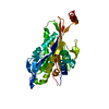
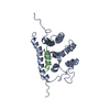
- PDB-1sdm: Crystal structure of kinesin-like calmodulin binding protein -

+
Open data


ID or keywords:

Loading...

-
Basic information

Entry
Database: PDB / ID: 1sdm
TitleCrystal structure of kinesin-like calmodulin binding protein
Componentskinesin heavy chain-like protein
KeywordsTRANSPORT PROTEIN / kinesin / minus-end directed / Ca2+/calmodulin regulated
Function / homology
Function and homology information


microtubule motor activity / microtubule-based movement / microtubule binding / oxidoreductase activity / cytoskeleton / ATP binding / metal ion binding
Similarity search - Function
MyTH4 domain / MyTH4 domain superfamily / MyTH4 domain / MyTH4 domain profile. / Domain in Myosin and Kinesin Tails / Prismane-like superfamily / Kinesin motor domain / Kinesin / Kinesin-like protein / FERM central domain ...MyTH4 domain / MyTH4 domain superfamily / MyTH4 domain / MyTH4 domain profile. / Domain in Myosin and Kinesin Tails / Prismane-like superfamily / Kinesin motor domain / Kinesin / Kinesin-like protein / FERM central domain / Kinesin motor domain signature. / Kinesin motor domain, conserved site / Kinesin motor domain / Kinesin motor domain profile. / Kinesin motor, catalytic domain. ATPase. / Kinesin motor domain / FERM/acyl-CoA-binding protein superfamily / FERM central domain / FERM superfamily, second domain / FERM domain / FERM domain profile. / Band 4.1 domain / Band 4.1 homologues / Kinesin motor domain superfamily / PH-like domain superfamily / P-loop containing nucleoside triphosphate hydrolase / 3-Layer(aba) Sandwich / Alpha Beta
Similarity search - Domain/homology
ADENOSINE-5'-DIPHOSPHATE / Kinesin heavy chain
Similarity search - Component
Biological speciesSolanum tuberosum (potato)
MethodX-RAY DIFFRACTION / SYNCHROTRON / MOLECULAR REPLACEMENT / Resolution: 2.3 Å
AuthorsVinogradova, M.V. / Reddy, V.S. / Reddy, A.S. / Sablin, E.P. / Fletterick, R.J.
CitationJournal: J.Biol.Chem. / Year: 2004
Title: Crystal structure of kinesin regulated by Ca(2+)-calmodulin.
Authors: Vinogradova, M.V. / Reddy, V.S. / Reddy, A.S. / Sablin, E.P. / Fletterick, R.J.
History
DepositionFeb 13, 2004Deposition site: RCSB / Processing site: RCSB
Revision 1.0Jun 22, 2004Provider: repository / Type: Initial release
Revision 1.1Apr 29, 2008Group: Version format compliance
Revision 1.2Jul 13, 2011Group: Version format compliance
Revision 1.3Aug 23, 2023Group: Data collection / Database references ...Data collection / Database references / Derived calculations / Refinement description
Category: chem_comp_atom / chem_comp_bond ...chem_comp_atom / chem_comp_bond / database_2 / pdbx_initial_refinement_model / pdbx_struct_conn_angle / struct_conn / struct_site
Item: _database_2.pdbx_DOI / _database_2.pdbx_database_accession ..._database_2.pdbx_DOI / _database_2.pdbx_database_accession / _pdbx_struct_conn_angle.ptnr1_auth_comp_id / _pdbx_struct_conn_angle.ptnr1_auth_seq_id / _pdbx_struct_conn_angle.ptnr1_label_asym_id / _pdbx_struct_conn_angle.ptnr1_label_atom_id / _pdbx_struct_conn_angle.ptnr1_label_comp_id / _pdbx_struct_conn_angle.ptnr1_label_seq_id / _pdbx_struct_conn_angle.ptnr3_auth_comp_id / _pdbx_struct_conn_angle.ptnr3_auth_seq_id / _pdbx_struct_conn_angle.ptnr3_label_asym_id / _pdbx_struct_conn_angle.ptnr3_label_atom_id / _pdbx_struct_conn_angle.ptnr3_label_comp_id / _pdbx_struct_conn_angle.ptnr3_label_seq_id / _pdbx_struct_conn_angle.value / _struct_conn.pdbx_dist_value / _struct_conn.ptnr1_auth_comp_id / _struct_conn.ptnr1_auth_seq_id / _struct_conn.ptnr1_label_asym_id / _struct_conn.ptnr1_label_atom_id / _struct_conn.ptnr1_label_comp_id / _struct_conn.ptnr2_auth_comp_id / _struct_conn.ptnr2_auth_seq_id / _struct_conn.ptnr2_label_asym_id / _struct_conn.ptnr2_label_atom_id / _struct_conn.ptnr2_label_comp_id / _struct_conn.ptnr2_label_seq_id / _struct_site.pdbx_auth_asym_id / _struct_site.pdbx_auth_comp_id / _struct_site.pdbx_auth_seq_id

-
Structure visualization

Structure viewerMolecule:
MolmilJmol/JSmol

Downloads & links

-
Assembly

Deposited unit
A: kinesin heavy chain-like protein
hetero molecules


Theoretical massNumber of molelcules
Total (without water)41,8803
Polymers41,4291
Non-polymers4522
Water2,018112
1


  • Idetical with deposited unit
  • defined by author
TypeNameSymmetry operationNumber
identity operation1_555x,y,z1
Unit cell
Length a, b, c (Å)95.728, 85.302, 44.457
Angle α, β, γ (deg.)90.00, 90.00, 90.00
Int Tables number18
Space group name H-MP21212

-
Components

#1: Protein kinesin heavy chain-like protein / KINESIN-LIKE CALMODULIN BINDING PROTEIN


Mass: 41428.910 Da / Num. of mol.: 1
Fragment: motor domain with adjacent C-terminal regulatory domain
Source method: isolated from a genetically manipulated source
Source: (gene. exp.) Solanum tuberosum (potato) / Production host: Escherichia coli (E. coli) / References: UniProt: Q41460
#2: Chemical ChemComp-MG / MAGNESIUM ION


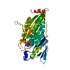
Mass: 24.305 Da / Num. of mol.: 1 / Source method: obtained synthetically / Formula: Mg
#3: Chemical ChemComp-ADP / ADENOSINE-5'-DIPHOSPHATE


Mass: 427.201 Da / Num. of mol.: 1 / Source method: obtained synthetically / Formula: C10H15N5O10P2 / Comment: ADP, energy-carrying molecule*YM
#4: Water ChemComp-HOH / water


Mass: 18.015 Da / Num. of mol.: 112 / Source method: isolated from a natural source / Formula: H2O

-
Experimental details

-
Experiment

ExperimentMethod: X-RAY DIFFRACTION / Number of used crystals: 1

-
Sample preparation

CrystalDensity Matthews: 2.36 Å3/Da / Density % sol: 47.45 %
Crystal growTemperature: 277 K / Method: vapor diffusion, sitting drop / pH: 9.1
Details: 20% PEG3350, 0.2 M di-Sodium phosphate, pH 9.1, VAPOR DIFFUSION, SITTING DROP, temperature 277K

-
Data collection

DiffractionMean temperature: 200 K
Diffraction sourceSource: SYNCHROTRON / Site: ALS / Beamline: 8.3.1 / Wavelength: 1.1 Å
DetectorType: ADSC QUANTUM 4 / Detector: CCD / Date: May 9, 2003
RadiationMonochromator: double crystal / Protocol: SINGLE WAVELENGTH / Monochromatic (M) / Laue (L): M / Scattering type: x-ray
Radiation wavelengthWavelength: 1.1 Å / Relative weight: 1
ReflectionResolution: 2.3→25 Å / Num. all: 16976 / Num. obs: 15295 / % possible obs: 90.1 % / Observed criterion σ(F): 0 / Observed criterion σ(I): 0 / Redundancy: 3.3 % / Biso Wilson estimate: 19.2 Å2 / Rmerge(I) obs: 0.051 / Rsym value: 0.047 / Net I/σ(I): 25.6
Reflection shellResolution: 2.3→2.38 Å / Redundancy: 2.6 % / Rmerge(I) obs: 0.173 / Mean I/σ(I) obs: 5.5 / Num. unique all: 1284 / Rsym value: 0.2 / % possible all: 77.6

-
Processing

Software
NameVersionClassification
CNS1.1refinement
DENZOdata reduction
SCALEPACKdata scaling
CNS1.1phasing
RefinementMethod to determine structure: MOLECULAR REPLACEMENT
Starting model: PDB ENTRY 1MKJ
Resolution: 2.3→24.8 Å / Rfactor Rfree error: 0.009 / Data cutoff high absF: 360408.75 / Data cutoff low absF: 0 / Isotropic thermal model: OVERALL / Cross valid method: THROUGHOUT / σ(F): 0 / Stereochemistry target values: Engh & Huber
RfactorNum. reflection% reflectionSelection details
Rfree0.254 747 4.9 %RANDOM
Rwork0.215 ---
all0.219 15165 --
obs0.215 15165 90.3 %-
Solvent computationSolvent model: FLAT MODEL / Bsol: 31.4998 Å2 / ksol: 0.314653 e/Å3
Displacement parametersBiso mean: 34.2 Å2
Baniso -1Baniso -2Baniso -3
1-10.24 Å20 Å20 Å2
2---6.88 Å20 Å2
3----3.36 Å2
Refine analyze
FreeObs
Luzzati coordinate error0.32 Å0.27 Å
Luzzati d res low-5 Å
Luzzati sigma a0.28 Å0.23 Å
Refinement stepCycle: LAST / Resolution: 2.3→24.8 Å
ProteinNucleic acidLigandSolventTotal
Num. atoms2720 0 28 112 2860
Refine LS restraints
Refine-IDTypeDev ideal
X-RAY DIFFRACTIONc_bond_d0.008
X-RAY DIFFRACTIONc_angle_deg1.4
X-RAY DIFFRACTIONc_dihedral_angle_d24.6
X-RAY DIFFRACTIONc_improper_angle_d1.38
LS refinement shellResolution: 2.3→2.44 Å / Rfactor Rfree error: 0.029 / Total num. of bins used: 6
RfactorNum. reflection% reflection
Rfree0.291 100 4.7 %
Rwork0.24 2032 -
obs--78 %
Xplor file
Refine-IDSerial noParam fileTopol file
X-RAY DIFFRACTION1PROTEIN_REP.PARAMPROTEIN.TOP
X-RAY DIFFRACTION2WATER_REP.PARAMWATER.TOP
X-RAY DIFFRACTION3ADP_NEW.PARAMADP_NEW.TOP
X-RAY DIFFRACTION4ION.PARAMION.TOP

+
About Yorodumi

-
News

-
Feb 9, 2022. New format data for meta-information of EMDB entries

New format data for meta-information of EMDB entries

  • Version 3 of the EMDB header file is now the official format.
  • The previous official version 1.9 will be removed from the archive.

Related info.:EMDB header

External links:wwPDB to switch to version 3 of the EMDB data model

-
Aug 12, 2020. Covid-19 info

Covid-19 info

URL: https://pdbjlvh1.pdbj.org/emnavi/covid19.php

New page: Covid-19 featured information page in EM Navigator.

Related info.:Covid-19 info / Mar 5, 2020. Novel coronavirus structure data

+
Mar 5, 2020. Novel coronavirus structure data

Novel coronavirus structure data

Related info.:Yorodumi Speices / Aug 12, 2020. Covid-19 info

External links:COVID-19 featured content - PDBj / Molecule of the Month (242):Coronavirus Proteases

+
Jan 31, 2019. EMDB accession codes are about to change! (news from PDBe EMDB page)

EMDB accession codes are about to change! (news from PDBe EMDB page)

  • The allocation of 4 digits for EMDB accession codes will soon come to an end. Whilst these codes will remain in use, new EMDB accession codes will include an additional digit and will expand incrementally as the available range of codes is exhausted. The current 4-digit format prefixed with “EMD-” (i.e. EMD-XXXX) will advance to a 5-digit format (i.e. EMD-XXXXX), and so on. It is currently estimated that the 4-digit codes will be depleted around Spring 2019, at which point the 5-digit format will come into force.
  • The EM Navigator/Yorodumi systems omit the EMD- prefix.

Related info.:Q: What is EMD? / ID/Accession-code notation in Yorodumi/EM Navigator

External links:EMDB Accession Codes are Changing Soon! / Contact to PDBj

+
Jul 12, 2017. Major update of PDB

Major update of PDB

  • wwPDB released updated PDB data conforming to the new PDBx/mmCIF dictionary.
  • This is a major update changing the version number from 4 to 5, and with Remediation, in which all the entries are updated.
  • In this update, many items about electron microscopy experimental information are reorganized (e.g. em_software).
  • Now, EM Navigator and Yorodumi are based on the updated data.

External links:wwPDB Remediation / Enriched Model Files Conforming to OneDep Data Standards Now Available in the PDB FTP Archive

-
Yorodumi

Thousand views of thousand structures

  • Yorodumi is a browser for structure data from EMDB, PDB, SASBDB, etc.
  • This page is also the successor to EM Navigator detail page, and also detail information page/front-end page for Omokage search.
  • The word "yorodu" (or yorozu) is an old Japanese word meaning "ten thousand". "mi" (miru) is to see.

Related info.:EMDB / PDB / SASBDB / Comparison of 3 databanks / Yorodumi Search / Aug 31, 2016. New EM Navigator & Yorodumi / Yorodumi Papers / Jmol/JSmol / Function and homology information / Changes in new EM Navigator and Yorodumi

Read more