 Movie
Movie Controller


+ Open data

- Basic information
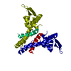
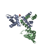
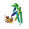
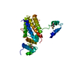
| Entry | Database: PDB / ID: 1sd6 | ||||||
|---|---|---|---|---|---|---|---|
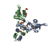
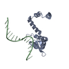
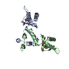
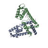
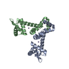
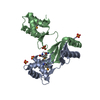
| Title | Crystal Structure of Native MecI at 2.65 A | ||||||
 Components Components | Methicillin resistance regulatory protein mecI | ||||||
 Keywords Keywords | DNA BINDING PROTEIN / BlaI / MecI / repressor / methicillin / B-lactam | ||||||
| Function / homology |  Function and homology information Function and homology informationresponse to antibiotic / negative regulation of DNA-templated transcription / DNA-templated transcription / DNA binding / cytoplasm Similarity search - Function | ||||||
| Biological species |   Staphylococcus aureus (bacteria) Staphylococcus aureus (bacteria) | ||||||
| Method |  X-RAY DIFFRACTION / FOURIER SYNTHESIS / Resolution: 2.65 Å X-RAY DIFFRACTION / FOURIER SYNTHESIS / Resolution: 2.65 Å | ||||||
 Authors Authors | Safo, M.K. / Zhao, Q. / Musayev, F.N. / Robinson, H. / Scarsdale, N. / Archer, G.L. | ||||||
 Citation Citation | Journal: J.Bacteriol. / Year: 2005 Title: Crystal structures of the BlaI repressor from Staphylococcus aureus and its complex with DNA: insights into transcriptional regulation of the bla and mec operons Authors: Safo, M.K. / Zhao, Q. / Ko, T.-P. / Musayev, F.N. / Robinson, H. / Scarsdale, N. / Wang, A.H.-J. / Archer, G.L. | ||||||
| History |
|
- Structure visualization
| Structure viewer | Molecule: MolmilJmol/JSmol |
|---|
- Downloads & links
-Download
| PDBx/mmCIF format | 1sd6.cif.gz | 60.8 KB | Display | PDBx/mmCIF format |
|---|---|---|---|---|
| PDB format | pdb1sd6.ent.gz | 45.8 KB | Display | PDB format |
| PDBx/mmJSON format | 1sd6.json.gz | Tree view | PDBx/mmJSON format | |
| Others |  Other downloads Other downloads |
-Validation report
| Arichive directory | https://data.pdbj.org/pub/pdb/validation_reports/sd/1sd6ftp://data.pdbj.org/pub/pdb/validation_reports/sd/1sd6 | HTTPS FTP |
|---|
-Related structure data

-Links
 PDBj
PDBj- Assembly
| Deposited unit | 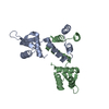
| ||||||||
|---|---|---|---|---|---|---|---|---|---|
| 1 |
| ||||||||
| Unit cell |
| ||||||||
| Details | The asymmetric unit contains the biological homodimer |
-Components
| #1: Protein | Mass: 14826.030 Da / Num. of mol.: 2 Source method: isolated from a genetically manipulated source Source: (gene. exp.) Staphylococcus aureus (bacteria) / Gene: MECI, SA0040 / Plasmid: pET3 / Production host: #2: Water | ChemComp-HOH / |  Mass: 18.015 Da / Num. of mol.: 59 / Source method: isolated from a natural source / Formula: H2O Mass: 18.015 Da / Num. of mol.: 59 / Source method: isolated from a natural source / Formula: H2O |
|---|
-Experimental details
-Experiment
| Experiment | Method: X-RAY DIFFRACTION / Number of used crystals: 1 |
|---|
- Sample preparation
| Crystal | Density Matthews: 2.88 Å3/Da / Density % sol: 57.23 % | |||||||||||||||||||||
|---|---|---|---|---|---|---|---|---|---|---|---|---|---|---|---|---|---|---|---|---|---|---|
| Crystal grow | Temperature: 291 K / Method: vapor diffusion, hanging drop / pH: 6.5 Details: PEG 8000, Sodium Cacodylate, Magnesium acetate , pH 6.5, VAPOR DIFFUSION, HANGING DROP, temperature 291K | |||||||||||||||||||||
| Crystal grow | *PLUS pH: 7.6 / Method: vapor diffusion, hanging drop | |||||||||||||||||||||
| Components of the solutions | *PLUS
|
-Data collection
| Diffraction | Mean temperature: 100 K |
|---|---|
| Diffraction source | Source: ROTATING ANODE / Type: RIGAKU RU200 / Wavelength: 1.5418 Å |
| Detector | Type: RIGAKU RAXIS II / Detector: IMAGE PLATE / Date: Mar 12, 2001 / Details: mirrors |
| Radiation | Monochromator: MSC Confocal Mirror / Protocol: SINGLE WAVELENGTH / Monochromatic (M) / Laue (L): M / Scattering type: x-ray |
| Radiation wavelength | Wavelength: 1.5418 Å / Relative weight: 1 |
| Reflection | Resolution: 2.65→36.48 Å / Num. all: 9536 / Num. obs: 9418 / % possible obs: 91.6 % / Observed criterion σ(F): 0 / Observed criterion σ(I): 0 / Redundancy: 2.8 % / Biso Wilson estimate: 35.9 Å2 / Rmerge(I) obs: 0.066 / Net I/σ(I): 10.2 |
| Reflection shell | Resolution: 2.65→2.78 Å / Redundancy: 2.9 % / Rmerge(I) obs: 0.37 / Mean I/σ(I) obs: 1.4 / Num. unique all: 1180 / % possible all: 88.4 |
- Processing
| Software |
| |||||||||||||||||||||||||
|---|---|---|---|---|---|---|---|---|---|---|---|---|---|---|---|---|---|---|---|---|---|---|---|---|---|---|
| Refinement | Method to determine structure: FOURIER SYNTHESIS / Resolution: 2.65→32.61 Å / Rfactor Rfree error: 0.012 / Data cutoff high absF: 91660.17 / Data cutoff low absF: 0 / Isotropic thermal model: RESTRAINED / Cross valid method: THROUGHOUT / σ(F): 0.5 / Stereochemistry target values: Engh & Huber
| |||||||||||||||||||||||||
| Solvent computation | Solvent model: FLAT MODEL / Bsol: 48.1141 Å2 / ksol: 0.311292 e/Å3 | |||||||||||||||||||||||||
| Displacement parameters | Biso mean: 71.4 Å2
| |||||||||||||||||||||||||
| Refine analyze |
| |||||||||||||||||||||||||
| Refinement step | Cycle: LAST / Resolution: 2.65→32.61 Å
| |||||||||||||||||||||||||
| Refine LS restraints |
| |||||||||||||||||||||||||
| LS refinement shell | Resolution: 2.65→2.82 Å / Rfactor Rfree error: 0.044 / Total num. of bins used: 6
| |||||||||||||||||||||||||
| Refine LS restraints | *PLUS
|
