 Movie
Movie Controller


+ Open data

- Basic information
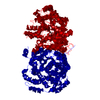
| Entry | Database: PDB / ID: 1s3h | ||||||
|---|---|---|---|---|---|---|---|
| Title | Propionibacterium shermanii transcarboxylase 5S subunit A59T | ||||||
 Components Components | transcarboxylase 5S subunit | ||||||
 Keywords Keywords | TRANSFERASE / TIM-barrel / carbamylated lysine / transcarboxylase / cobalt | ||||||
| Function / homology |  Function and homology information Function and homology informationmethylmalonyl-CoA carboxytransferase / methylmalonyl-CoA carboxytransferase activity / pyruvate carboxylase activity / gluconeogenesis / metal ion binding / cytoplasm Similarity search - Function | ||||||
| Biological species |  Propionibacterium freudenreichii subsp. shermanii (bacteria) Propionibacterium freudenreichii subsp. shermanii (bacteria) | ||||||
| Method |  X-RAY DIFFRACTION / Isomorphous replacement / Resolution: 2.5 Å X-RAY DIFFRACTION / Isomorphous replacement / Resolution: 2.5 Å | ||||||
 Authors Authors | Hall, P.R. / Zheng, R. / Antony, L. / Pusztai-Carey, M. / Carey, P.R. / Yee, V.C. | ||||||
 Citation Citation | Journal: Embo J. / Year: 2004 Title: Transcarboxylase 5S structures: assembly and catalytic mechanism of a multienzyme complex subunit. Authors: Hall, P.R. / Zheng, R. / Antony, L. / Pusztai-Carey, M. / Carey, P.R. / Yee, V.C. #1: Journal: ACTA CRYSTALLOGR.,SECT.D / Year: 2004 Title: Expression and crystallization of several forms of the Propionibacterium shermanii transcarboxylase 5S subunit. Authors: Hall, P.R. / Zheng, R. / Pusztai-Carey, M. / van den Akker, F. / Carey, P.R. / Yee, V.C. | ||||||
| History |
|
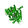
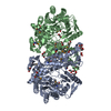
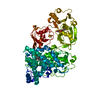
- Structure visualization
| Structure viewer | Molecule: MolmilJmol/JSmol |
|---|
- Downloads & links
-Download
| PDBx/mmCIF format | 1s3h.cif.gz | 105.6 KB | Display | PDBx/mmCIF format |
|---|---|---|---|---|
| PDB format | pdb1s3h.ent.gz | 78.6 KB | Display | PDB format |
| PDBx/mmJSON format | 1s3h.json.gz | Tree view | PDBx/mmJSON format | |
| Others |  Other downloads Other downloads |
-Validation report
| Arichive directory | https://data.pdbj.org/pub/pdb/validation_reports/s3/1s3hftp://data.pdbj.org/pub/pdb/validation_reports/s3/1s3h | HTTPS FTP |
|---|
-Related structure data
| Related structure data | 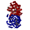 1rqbSC  1rqeC 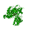 1rqhC 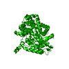 1rr2C  1u5jC  1s27 S: Starting model for refinement C: citing same article ( |
|---|---|
| Similar structure data |


-Links
 PDBj
PDBj
- Assembly
| Deposited unit | 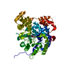
| ||||||||
|---|---|---|---|---|---|---|---|---|---|
| 1 | 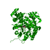
| ||||||||
| Unit cell |
| ||||||||
| Details | The asymmetric unit contains a monomer. The biological dimer is formed by the following transformation: -1.0000*x+ 0.0000*y+ 0.0000*z+ 192.516 0.0000*x+ 1.0000*y+ 0.0000*z+ 0.000 0.0000*x+ 0.0000*y+ -1.0000*z+ 39.359 |
-Components
| #1: Protein | Mass: 59682.496 Da / Num. of mol.: 1 / Mutation: A59T Source method: isolated from a genetically manipulated source Source: (gene. exp.) Propionibacterium freudenreichii subsp. shermanii (bacteria)Species: Propionibacterium freudenreichii / Strain: subsp. shermanii / Gene: 5S / Plasmid: pETBlue-2 / Production host: References: UniProt: Q70AC7, methylmalonyl-CoA carboxytransferase |
|---|---|
| #2: Chemical | ChemComp-CO /   Mass: 58.933 Da / Num. of mol.: 1 / Source method: obtained synthetically / Formula: Co Mass: 58.933 Da / Num. of mol.: 1 / Source method: obtained synthetically / Formula: Co |
| #3: Water | ChemComp-HOH /  Mass: 18.015 Da / Num. of mol.: 1 / Source method: isolated from a natural source / Formula: H2O Mass: 18.015 Da / Num. of mol.: 1 / Source method: isolated from a natural source / Formula: H2O |
-Experimental details
-Experiment
| Experiment | Method: X-RAY DIFFRACTION / Number of used crystals: 1 |
|---|
- Sample preparation
| Crystal | Density Matthews: 2.32 Å3/Da / Density % sol: 47.03 % |
|---|---|
| Crystal grow | Temperature: 293 K / Method: vapor diffusion, sitting drop / pH: 7 Details: PEG 4000, Tris , pH 7.0, VAPOR DIFFUSION, SITTING DROP, temperature 293K |
-Data collection
| Diffraction | Mean temperature: 95 K |
|---|---|
| Diffraction source | Source: ROTATING ANODE / Type: RIGAKU RUH3R / Wavelength: 1.5418 |
| Detector | Type: RIGAKU RAXIS IV / Detector: IMAGE PLATE / Date: Jun 23, 2003 |
| Radiation | Monochromator: Yale mirrors / Protocol: SINGLE WAVELENGTH / Monochromatic (M) / Laue (L): M / Scattering type: x-ray |
| Radiation wavelength | Wavelength: 1.5418 Å / Relative weight: 1 |
| Reflection | Resolution: 2.5→30 Å / Num. all: 19598 / Num. obs: 18621 / % possible obs: 95 % / Observed criterion σ(F): 0 / Observed criterion σ(I): 0 / Redundancy: 7.6 % / Biso Wilson estimate: 22.7 Å2 / Rmerge(I) obs: 0.069 / Net I/σ(I): 22 |
| Reflection shell | Resolution: 2.5→2.59 Å / Rmerge(I) obs: 0.177 / Mean I/σ(I) obs: 6.9 / % possible all: 87.5 |
- Processing
| Software |
| |||||||||||||||||||||||||
|---|---|---|---|---|---|---|---|---|---|---|---|---|---|---|---|---|---|---|---|---|---|---|---|---|---|---|
| Refinement | Method to determine structure: Isomorphous replacement Starting model: PDB ENTRY 1RQB Resolution: 2.5→29.19 Å / Rfactor Rfree error: 0.008 / Data cutoff high absF: 226821.14 / Data cutoff low absF: 0 / Isotropic thermal model: RESTRAINED / Cross valid method: THROUGHOUT / σ(F): 0 / Stereochemistry target values: Engh & Huber
| |||||||||||||||||||||||||
| Solvent computation | Solvent model: FLAT MODEL / Bsol: 10 Å2 / ksol: 0.300775 e/Å3 | |||||||||||||||||||||||||
| Displacement parameters | Biso mean: 20.2 Å2
| |||||||||||||||||||||||||
| Refine analyze |
| |||||||||||||||||||||||||
| Refinement step | Cycle: LAST / Resolution: 2.5→29.19 Å
| |||||||||||||||||||||||||
| Refine LS restraints |
| |||||||||||||||||||||||||
| LS refinement shell | Resolution: 2.5→2.66 Å / Rfactor Rfree error: 0.024 / Total num. of bins used: 6
| |||||||||||||||||||||||||
| Xplor file |
|
