 Movie
Movie Controller

[English] 日本語
 Yorodumi
- PDB-1rzv: Crystal structure of the glycogen synthase from Agrobacterium tum... -
+ Open data

- Basic information
| Entry | Database: PDB / ID: 1rzv | ||||||
|---|---|---|---|---|---|---|---|
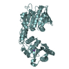
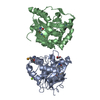
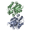
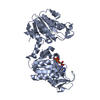
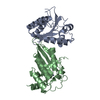
| Title | Crystal structure of the glycogen synthase from Agrobacterium tumefaciens (non-complexed form) | ||||||
 Components Components | Glycogen synthase 1 | ||||||
 Keywords Keywords | TRANSFERASE / glycosyl-transferase GT-B fold / Rossmann fold | ||||||
| Function / homology |  Function and homology information Function and homology informationstarch synthase (glycosyl-transferring) / alpha-1,4-glucan glucosyltransferase (ADP-glucose donor) activity / alpha-1,4-glucan glucosyltransferase (UDP-glucose donor) activity / glycogen biosynthetic process / cytosol Similarity search - Function | ||||||
| Biological species |  Agrobacterium tumefaciens (bacteria) Agrobacterium tumefaciens (bacteria) | ||||||
| Method |  X-RAY DIFFRACTION / SYNCHROTRON / SAD / Resolution: 2.3 Å X-RAY DIFFRACTION / SYNCHROTRON / SAD / Resolution: 2.3 Å | ||||||
 Authors Authors | Buschiazzo, A. / Guerin, M.E. / Ugalde, J.E. / Ugalde, R.A. / Shepard, W. / Alzari, P.M. | ||||||
 Citation Citation | Journal: Embo J. / Year: 2004 Title: Crystal structure of glycogen synthase: homologous enzymes catalyze glycogen synthesis and degradation. Authors: Buschiazzo, A. / Ugalde, J.E. / Guerin, M.E. / Shepard, W. / Ugalde, R.A. / Alzari, P.M. #1: Journal: Acta Crystallogr.,Sect.D / Year: 2003Title: Preliminary crystallographic studies of glycogen synthase from Agrobacterium tumefaciens Authors: Guerin, M.E. / Buschiazzo, A. / Ugalde, J.E. / Ugalde, R.A. / Alzari, P.M. | ||||||
| History |
|
- Structure visualization
| Structure viewer | Molecule: MolmilJmol/JSmol |
|---|
- Downloads & links
-Download
| PDBx/mmCIF format | 1rzv.cif.gz | 197 KB | Display | PDBx/mmCIF format |
|---|---|---|---|---|
| PDB format | pdb1rzv.ent.gz | 157.7 KB | Display | PDB format |
| PDBx/mmJSON format | 1rzv.json.gz | Tree view | PDBx/mmJSON format | |
| Others |  Other downloads Other downloads |
-Validation report
| Arichive directory | https://data.pdbj.org/pub/pdb/validation_reports/rz/1rzvftp://data.pdbj.org/pub/pdb/validation_reports/rz/1rzv | HTTPS FTP |
|---|
-Related structure data


-Links
 PDBj
PDBj
- Assembly
| Deposited unit | 
| ||||||||
|---|---|---|---|---|---|---|---|---|---|
| 1 | 
| ||||||||
| 2 | 
| ||||||||
| Unit cell |
|
-Components
| #1: Protein | Mass: 52925.734 Da / Num. of mol.: 2 Source method: isolated from a genetically manipulated source Source: (gene. exp.) Agrobacterium tumefaciens (bacteria) / Gene: GLGA1, GLGA, ATU4075, AGR_L_1562 / Plasmid: pBBRMCS-4 / Production host: Agrobacterium tumefaciens (bacteria) / Strain (production host): A5130References: UniProt: P0A3F3, starch synthase (glycosyl-transferring) #2: Water | ChemComp-HOH / |  Mass: 18.015 Da / Num. of mol.: 407 / Source method: isolated from a natural source / Formula: H2O Mass: 18.015 Da / Num. of mol.: 407 / Source method: isolated from a natural source / Formula: H2OHas protein modification | Y | |
|---|
-Experimental details
-Experiment
| Experiment | Method: X-RAY DIFFRACTION / Number of used crystals: 1 |
|---|
- Sample preparation
| Crystal | Density Matthews: 2.6 Å3/Da / Density % sol: 52.24 % |
|---|---|
| Crystal grow | Temperature: 291 K / Method: vapor diffusion, hanging drop / pH: 7.5 Details: PEG 4000, isopropanol, HEPES, pH 7.5, VAPOR DIFFUSION, HANGING DROP, temperature 291K |
-Data collection
| Diffraction | Mean temperature: 110 K |
|---|---|
| Diffraction source | Source: SYNCHROTRON / Site: ESRF  / Beamline: ID29 / Wavelength: 0.9792 Å / Beamline: ID29 / Wavelength: 0.9792 Å |
| Detector | Type: ADSC QUANTUM 210 / Detector: CCD / Date: Nov 11, 2002 Details: channel - cut Si monochromator + cylindrical grazing incidence mirror |
| Radiation | Monochromator: channel cut Si(311) / Protocol: SINGLE WAVELENGTH / Monochromatic (M) / Laue (L): M / Scattering type: x-ray |
| Radiation wavelength | Wavelength: 0.9792 Å / Relative weight: 1 |
| Reflection | Resolution: 2.3→29.97 Å / Num. all: 88607 / Num. obs: 88607 / % possible obs: 98.2 % / Observed criterion σ(F): 0 / Observed criterion σ(I): 0 / Redundancy: 6.7 % / Biso Wilson estimate: 23 Å2 / Limit h max: 29 / Limit h min: 0 / Limit k max: 37 / Limit k min: 0 / Limit l max: 37 / Limit l min: -38 / Observed criterion F max: 1545415.8 / Observed criterion F min: 14.7 / Rmerge(I) obs: 0.072 / Rsym value: 0.055 / Net I/σ(I): 11.4 |
| Reflection shell | Resolution: 2.3→2.4 Å / Redundancy: 3.8 % / Rmerge(I) obs: 0.257 / Mean I/σ(I) obs: 5.5 / Num. unique all: 9708 / Rsym value: 0.188 / % possible all: 85.9 |
- Processing
| Software |
| ||||||||||||||||||||||||||||||||||||||||||||||||||||||||||||||||||||||||||||||||||||||||||
|---|---|---|---|---|---|---|---|---|---|---|---|---|---|---|---|---|---|---|---|---|---|---|---|---|---|---|---|---|---|---|---|---|---|---|---|---|---|---|---|---|---|---|---|---|---|---|---|---|---|---|---|---|---|---|---|---|---|---|---|---|---|---|---|---|---|---|---|---|---|---|---|---|---|---|---|---|---|---|---|---|---|---|---|---|---|---|---|---|---|---|---|
| Refinement | Method to determine structure: SAD / Resolution: 2.3→29.97 Å / Rfactor Rfree error: 0.004 / Occupancy max: 1 / Occupancy min: 1 / Cross valid method: THROUGHOUT / σ(F): 0 / σ(I): 0 / Stereochemistry target values: Engh & Huber
| ||||||||||||||||||||||||||||||||||||||||||||||||||||||||||||||||||||||||||||||||||||||||||
| Solvent computation | Solvent model: CNS bulk solvent model used / Bsol: 43.6171 Å2 / ksol: 0.380688 e/Å3 | ||||||||||||||||||||||||||||||||||||||||||||||||||||||||||||||||||||||||||||||||||||||||||
| Displacement parameters | Biso max: 77.94 Å2 / Biso mean: 31.42 Å2 / Biso min: 10.44 Å2
| ||||||||||||||||||||||||||||||||||||||||||||||||||||||||||||||||||||||||||||||||||||||||||
| Refine Biso |
| ||||||||||||||||||||||||||||||||||||||||||||||||||||||||||||||||||||||||||||||||||||||||||
| Refine analyze |
| ||||||||||||||||||||||||||||||||||||||||||||||||||||||||||||||||||||||||||||||||||||||||||
| Refinement step | Cycle: LAST / Resolution: 2.3→29.97 Å
| ||||||||||||||||||||||||||||||||||||||||||||||||||||||||||||||||||||||||||||||||||||||||||
| Refine LS restraints |
| ||||||||||||||||||||||||||||||||||||||||||||||||||||||||||||||||||||||||||||||||||||||||||
| LS refinement shell | Refine-ID: X-RAY DIFFRACTION
| ||||||||||||||||||||||||||||||||||||||||||||||||||||||||||||||||||||||||||||||||||||||||||
| Xplor file |
|
