 Movie
Movie Controller

[English] 日本語
 Yorodumi
- PDB-1r1k: Crystal structure of the ligand-binding domains of the heterodime... -
+ Open data

- Basic information
| Entry | Database: PDB / ID: 1r1k | ||||||
|---|---|---|---|---|---|---|---|
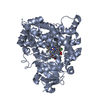
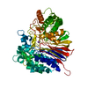
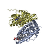
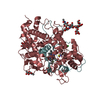
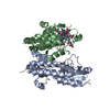
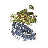
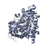
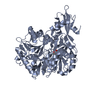
| Title | Crystal structure of the ligand-binding domains of the heterodimer EcR/USP bound to ponasterone A | ||||||
 Components Components |
| ||||||
 Keywords Keywords | hormone/growth factor receptor / NUCLEAR RECEPTOR / TRANSCRIPTION REGULATION / HETERODIMER / ALPHA-HELICAL SANDWICH / Structural Proteomics in Europe / SPINE / Structural Genomics / hormone-growth factor receptor COMPLEX | ||||||
| Function / homology |  Function and homology information Function and homology informationecdysone binding / ecdysone receptor signaling pathway / nuclear steroid receptor activity / RNA polymerase II transcription regulator complex / nuclear receptor activity / cell differentiation / RNA polymerase II cis-regulatory region sequence-specific DNA binding / negative regulation of transcription by RNA polymerase II / positive regulation of transcription by RNA polymerase II / DNA binding ...ecdysone binding / ecdysone receptor signaling pathway / nuclear steroid receptor activity / RNA polymerase II transcription regulator complex / nuclear receptor activity / cell differentiation / RNA polymerase II cis-regulatory region sequence-specific DNA binding / negative regulation of transcription by RNA polymerase II / positive regulation of transcription by RNA polymerase II / DNA binding / zinc ion binding / nucleus Similarity search - Function | ||||||
| Biological species |  Heliothis virescens (tobacco budworm) Heliothis virescens (tobacco budworm) | ||||||
| Method |  X-RAY DIFFRACTION / SYNCHROTRON / MOLECULAR REPLACEMENT / Resolution: 2.9 Å X-RAY DIFFRACTION / SYNCHROTRON / MOLECULAR REPLACEMENT / Resolution: 2.9 Å | ||||||
 Authors Authors | Billas, I.M.L. / Iwema, T. / Garnier, J.-M. / Mitschler, A. / Rochel, N. / Moras, D. / Structural Proteomics in Europe (SPINE) | ||||||
 Citation Citation | Journal: Nature / Year: 2003 Title: Structural adaptability in the ligand-binding pocket of the ecdysone hormone receptor. Authors: Billas, I.M.L. / Iwema, T. / Garnier, J.M. / Mitschler, A. / Rochel, N. / Moras, D. | ||||||
| History |
| ||||||
| Remark 999 | sequence no suitable sequence database reference was available for chains A or D, at the time of ...sequence no suitable sequence database reference was available for chains A or D, at the time of processing this file |
- Structure visualization
| Structure viewer | Molecule: MolmilJmol/JSmol |
|---|
- Downloads & links
-Download
| PDBx/mmCIF format | 1r1k.cif.gz | 113.7 KB | Display | PDBx/mmCIF format |
|---|---|---|---|---|
| PDB format | pdb1r1k.ent.gz | 85.1 KB | Display | PDB format |
| PDBx/mmJSON format | 1r1k.json.gz | Tree view | PDBx/mmJSON format | |
| Others |  Other downloads Other downloads |
-Validation report
| Summary document | 1r1k_validation.pdf.gz | 964.1 KB | Display | wwPDB validaton report |
|---|---|---|---|---|
| Full document | 1r1k_full_validation.pdf.gz | 995.2 KB | Display | |
| Data in XML | 1r1k_validation.xml.gz | 23.5 KB | Display | |
| Data in CIF | 1r1k_validation.cif.gz | 30.3 KB | Display | |
| Arichive directory | https://data.pdbj.org/pub/pdb/validation_reports/r1/1r1kftp://data.pdbj.org/pub/pdb/validation_reports/r1/1r1k | HTTPS FTP |
-Related structure data
| Related structure data |  1r20C 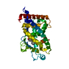 1db1S C: citing same article ( S: Starting model for refinement |
|---|---|
| Similar structure data | |
| Other databases |


-Links
 PDBj
PDBj

- Assembly
| Deposited unit | 
| ||||||||
|---|---|---|---|---|---|---|---|---|---|
| 1 |
| ||||||||
| 2 | 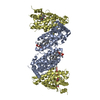
| ||||||||
| Unit cell |
|
-Components
| #1: Protein | Mass: 30368.961 Da / Num. of mol.: 1 / Fragment: hormone binding domain Source method: isolated from a genetically manipulated source Source: (gene. exp.) Heliothis virescens (tobacco budworm) / Gene: ECR OR NR1H1 / Plasmid: pET32b / Species (production host): Escherichia coli / Production host:  |
|---|---|
| #2: Protein | Mass: 29951.760 Da / Num. of mol.: 1 Source method: isolated from a genetically manipulated source Source: (gene. exp.) Heliothis virescens (tobacco budworm) / Plasmid: pACYC11b / Species (production host): Escherichia coli / Production host: |
| #3: Chemical | ChemComp-P1A /   Mass: 464.635 Da / Num. of mol.: 1 / Source method: obtained synthetically / Formula: C27H44O6 Mass: 464.635 Da / Num. of mol.: 1 / Source method: obtained synthetically / Formula: C27H44O6 |
| #4: Chemical | ChemComp-EPH /   Mass: 709.933 Da / Num. of mol.: 1 / Source method: obtained synthetically / Formula: C39H68NO8P / Comment: phospholipid*YM Mass: 709.933 Da / Num. of mol.: 1 / Source method: obtained synthetically / Formula: C39H68NO8P / Comment: phospholipid*YM |
| #5: Water | ChemComp-HOH /  Mass: 18.015 Da / Num. of mol.: 8 / Source method: isolated from a natural source / Formula: H2O Mass: 18.015 Da / Num. of mol.: 8 / Source method: isolated from a natural source / Formula: H2O |
-Experimental details
-Experiment
| Experiment | Method: X-RAY DIFFRACTION / Number of used crystals: 1 |
|---|
- Sample preparation
| Crystal | Density Matthews: 2.39 Å3/Da / Density % sol: 48.44 % | ||||||||||||||||||||||||||||||||||||||||||||||||||||||||||||||||||||||
|---|---|---|---|---|---|---|---|---|---|---|---|---|---|---|---|---|---|---|---|---|---|---|---|---|---|---|---|---|---|---|---|---|---|---|---|---|---|---|---|---|---|---|---|---|---|---|---|---|---|---|---|---|---|---|---|---|---|---|---|---|---|---|---|---|---|---|---|---|---|---|---|
| Crystal grow | Temperature: 298 K / Method: vapor diffusion, sitting drop / pH: 5.6 Details: PEG 4000, ammonium sulfate, pH 5.6, VAPOR DIFFUSION, SITTING DROP, temperature 298K | ||||||||||||||||||||||||||||||||||||||||||||||||||||||||||||||||||||||
| Crystal grow | *PLUS Temperature: 24 ℃ / pH: 8 / Method: vapor diffusion, hanging drop | ||||||||||||||||||||||||||||||||||||||||||||||||||||||||||||||||||||||
| Components of the solutions | *PLUS
|
-Data collection
| Diffraction | Mean temperature: 110 K |
|---|---|
| Diffraction source | Source: SYNCHROTRON / Site: ESRF  / Beamline: BM30A / Wavelength: 0.9798 Å / Beamline: BM30A / Wavelength: 0.9798 Å |
| Detector | Type: MARRESEARCH / Detector: CCD / Date: Sep 28, 2002 / Details: mirrors |
| Radiation | Monochromator: SAGITALLY FOCUSED Si(111) / Protocol: SINGLE WAVELENGTH / Monochromatic (M) / Laue (L): M / Scattering type: x-ray |
| Radiation wavelength | Wavelength: 0.9798 Å / Relative weight: 1 |
| Reflection | Resolution: 2.9→20 Å / Num. all: 13703 / Num. obs: 13703 / % possible obs: 99.5 % / Observed criterion σ(I): -3 / Rsym value: 0.052 |
| Reflection shell | Resolution: 2.9→2.97 Å / Rsym value: 0.38 / % possible all: 97.9 |
| Reflection | *PLUS Redundancy: 9.5 % / Num. measured all: 129598 / Rmerge(I) obs: 0.052 |
| Reflection shell | *PLUS % possible obs: 97.9 % / Rmerge(I) obs: 0.38 / Mean I/σ(I) obs: 4.2 |
- Processing
| Software |
| ||||||||||||||||||||
|---|---|---|---|---|---|---|---|---|---|---|---|---|---|---|---|---|---|---|---|---|---|
| Refinement | Method to determine structure: MOLECULAR REPLACEMENT Starting model: Vitamin D receptor, PDB entry 1DB1 Resolution: 2.9→20 Å / σ(F): 0 / Stereochemistry target values: Engh & Huber
| ||||||||||||||||||||
| Refinement step | Cycle: LAST / Resolution: 2.9→20 Å
| ||||||||||||||||||||
| Refine LS restraints |
| ||||||||||||||||||||
| Refinement | *PLUS % reflection Rfree: 10 % | ||||||||||||||||||||
| Solvent computation | *PLUS | ||||||||||||||||||||
| Displacement parameters | *PLUS | ||||||||||||||||||||
| Refine LS restraints | *PLUS
|
