[English] 日本語
Yorodumi
- PDB-1qz4: Structure of YcfC Protein of Unknown Function Escherichia coli -

+
Open data


ID or keywords:

Loading...

-
Basic information

Entry
Database: PDB / ID: 1qz4
TitleStructure of YcfC Protein of Unknown Function Escherichia coli
ComponentsHypothetical protein ycfCHypothesis
KeywordsStructural genomics / unknown function / helix-turn-helix motif / PSI / Protein Structure Initiative / Midwest Center for Structural Genomics / MCSG
Function / homology
Function and homology information


response to heat / plasma membrane / cytoplasm
Similarity search - Function
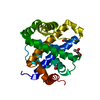
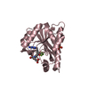
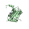
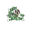
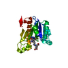
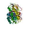
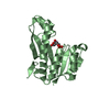
YcfC-like / HflD-like / High frequency lysogenization protein HflD / HflD-like superfamily / Protein of unknown function (DUF489) / Orthogonal Bundle / Mainly Alpha
Similarity search - Domain/homology
: / PHOSPHATE ION / High frequency lysogenization protein HflD
Similarity search - Component
Biological speciesEscherichia coli (E. coli)
MethodX-RAY DIFFRACTION / SYNCHROTRON / MIRAS / Resolution: 2 Å
AuthorsOtwinowski, Z. / Midwest Center for Structural Genomics (MCSG)
CitationJournal: To be Published
Title: Structure of a hypothetical protein ycfC coded by Escherichia coli genome.
Authors: Otwinowski, Z. / Borek, D.M. / Chen, Y. / Skarina, T. / Savchenko, A. / Edwards, A.
History
DepositionSep 15, 2003Deposition site: RCSB / Processing site: RCSB
Revision 1.0Mar 30, 2004Provider: repository / Type: Initial release
Revision 1.1Apr 29, 2008Group: Version format compliance
Revision 1.2Jul 13, 2011Group: Version format compliance
Revision 1.3Feb 14, 2024Group: Data collection / Database references / Derived calculations
Category: chem_comp_atom / chem_comp_bond ...chem_comp_atom / chem_comp_bond / database_2 / pdbx_struct_conn_angle / struct_conn / struct_ref_seq_dif / struct_site
Item: _database_2.pdbx_DOI / _database_2.pdbx_database_accession ..._database_2.pdbx_DOI / _database_2.pdbx_database_accession / _pdbx_struct_conn_angle.ptnr1_auth_comp_id / _pdbx_struct_conn_angle.ptnr1_auth_seq_id / _pdbx_struct_conn_angle.ptnr1_label_asym_id / _pdbx_struct_conn_angle.ptnr1_label_atom_id / _pdbx_struct_conn_angle.ptnr1_label_comp_id / _pdbx_struct_conn_angle.ptnr1_label_seq_id / _pdbx_struct_conn_angle.ptnr1_symmetry / _pdbx_struct_conn_angle.ptnr2_auth_seq_id / _pdbx_struct_conn_angle.ptnr2_label_asym_id / _pdbx_struct_conn_angle.ptnr3_auth_comp_id / _pdbx_struct_conn_angle.ptnr3_auth_seq_id / _pdbx_struct_conn_angle.ptnr3_label_asym_id / _pdbx_struct_conn_angle.ptnr3_label_atom_id / _pdbx_struct_conn_angle.ptnr3_label_comp_id / _pdbx_struct_conn_angle.ptnr3_label_seq_id / _pdbx_struct_conn_angle.ptnr3_symmetry / _pdbx_struct_conn_angle.value / _struct_conn.pdbx_dist_value / _struct_conn.ptnr1_auth_comp_id / _struct_conn.ptnr1_auth_seq_id / _struct_conn.ptnr1_label_asym_id / _struct_conn.ptnr1_label_atom_id / _struct_conn.ptnr1_label_comp_id / _struct_conn.ptnr1_label_seq_id / _struct_conn.ptnr1_symmetry / _struct_conn.ptnr2_auth_comp_id / _struct_conn.ptnr2_auth_seq_id / _struct_conn.ptnr2_label_asym_id / _struct_conn.ptnr2_label_atom_id / _struct_conn.ptnr2_label_comp_id / _struct_conn.ptnr2_label_seq_id / _struct_conn.ptnr2_symmetry / _struct_ref_seq_dif.details / _struct_site.pdbx_auth_asym_id / _struct_site.pdbx_auth_comp_id / _struct_site.pdbx_auth_seq_id
Revision 1.4Apr 3, 2024Group: Refinement description / Category: pdbx_initial_refinement_model

-
Structure visualization

Structure viewerMolecule:
MolmilJmol/JSmol

Downloads & links

-
Assembly

Deposited unit
A: Hypothetical protein ycfC
hetero molecules


Theoretical massNumber of molelcules
Total (without water)24,99812
Polymers22,8971
Non-polymers2,10111
Water2,000111
1


  • Idetical with deposited unit
  • defined by author
TypeNameSymmetry operationNumber
identity operation1_555x,y,z1
Unit cell
Length a, b, c (Å)43.102, 62.749, 68.399
Angle α, β, γ (deg.)90.00, 90.00, 90.00
Int Tables number19
Cell settingorthorhombic
Space group name H-MP212121

-
Components

#1: Protein Hypothetical protein ycfC / Hypothesis / ORF-23


Mass: 22897.371 Da / Num. of mol.: 1 / Fragment: ycfC protein / Mutation: none
Source method: isolated from a genetically manipulated source
Source: (gene. exp.) Escherichia coli (E. coli) / Gene: YCFC / Production host: Escherichia coli (E. coli) / References: UniProt: P25746
#2: Chemical ChemComp-PO4 / PHOSPHATE ION / Phosphate


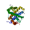
Mass: 94.971 Da / Num. of mol.: 1 / Source method: obtained synthetically / Formula: PO4
#3: Chemical
ChemComp-HG / MERCURY (II) ION / Mercury (element)


Mass: 200.590 Da / Num. of mol.: 10 / Source method: obtained synthetically / Formula: Hg
#4: Water ChemComp-HOH / water / Water


Mass: 18.015 Da / Num. of mol.: 111 / Source method: isolated from a natural source / Formula: H2O

-
Experimental details

-
Experiment

ExperimentMethod: X-RAY DIFFRACTION / Number of used crystals: 2

-
Sample preparation

CrystalDensity Matthews: 2.02 Å3/Da / Density % sol: 39.09 %
Crystal growTemperature: 293 K / Method: vapor diffusion, hanging drop
Details: PEG 3350, zinc acetate, lithium acetate , VAPOR DIFFUSION, HANGING DROP, temperature 293K

-
Data collection

Diffraction
IDMean temperature (K)Crystal-ID
11001
21001
Diffraction source
SourceSiteBeamlineIDWavelength (Å)
SYNCHROTRONAPS 19-ID10.999871
SYNCHROTRONAPS 19-ID20.91963
Detector
TypeIDDetectorDate
CUSTOM-MADE1CCDAug 24, 2003
CUSTOM-MADE2CCDJun 21, 2003
Radiation
IDMonochromatorProtocolMonochromatic (M) / Laue (L)Scattering typeWavelength-ID
1DOUBLE CRYSTAL MONOCHROMATOR SI-220SINGLE WAVELENGTHMx-ray1
2DOUBLE CRYSTAL MONOCHROMATOR SI-220SINGLE WAVELENGTHMx-ray1
Radiation wavelength
IDWavelength (Å)Relative weight
10.9998711
20.919631
ReflectionResolution: 1.95→50 Å / Num. all: 13895 / Num. obs: 11477 / % possible obs: 82.6 % / Observed criterion σ(F): 0 / Observed criterion σ(I): -3 / Rmerge(I) obs: 0.107 / Net I/σ(I): 15.87
Reflection shellResolution: 1.95→1.98 Å / Rmerge(I) obs: 0.423 / Mean I/σ(I) obs: 1.8 / % possible all: 36.7

-
Processing

Software
NameVersionClassification
REFMAC5.1.24refinement
DENZOdata reduction
SCALEPACKdata scaling
MLPHAREphasing
RefinementMethod to determine structure: MIRAS
Starting model: Build by WARP/ARP

Resolution: 2→25 Å / Cor.coef. Fo:Fc: 0.947 / SU B: 2.014 / SU ML: 0.058 / σ(F): 0 / ESU R: 0.212 / Details: HYDROGENS HAVE BEEN ADDED IN THE RIDING POSITIONS
RfactorNum. reflection% reflection
all0.15729 11138 -
obs0.15729 11138 85.24 %
Solvent computationIon probe radii: 0.8 Å / Shrinkage radii: 0.8 Å / VDW probe radii: 1.4 Å / Solvent model: BABINET MODEL WITH MASK
Displacement parametersBiso mean: 16.821 Å2
Baniso -1Baniso -2Baniso -3
1--0.99 Å20 Å20 Å2
2--0.8 Å20 Å2
3---0.19 Å2
Refinement stepCycle: LAST / Resolution: 2→25 Å
ProteinNucleic acidLigandSolventTotal
Num. atoms1605 0 15 111 1731
Refine LS restraints
Refine-IDTypeDev idealDev ideal targetNumber
X-RAY DIFFRACTIONr_bond_refined_d0.0180.0211620
X-RAY DIFFRACTIONr_bond_other_d0.0020.021543
X-RAY DIFFRACTIONr_angle_refined_deg1.5821.9742197
X-RAY DIFFRACTIONr_angle_other_deg0.91133567
X-RAY DIFFRACTIONr_dihedral_angle_1_deg5.2685212
X-RAY DIFFRACTIONr_chiral_restr0.0910.2267
X-RAY DIFFRACTIONr_gen_planes_refined0.0060.021805
X-RAY DIFFRACTIONr_gen_planes_other0.0030.02302
X-RAY DIFFRACTIONr_nbd_refined0.230.2390
X-RAY DIFFRACTIONr_nbd_other0.2310.21704
X-RAY DIFFRACTIONr_nbtor_other0.0860.2976
X-RAY DIFFRACTIONr_xyhbond_nbd_refined0.220.282
X-RAY DIFFRACTIONr_metal_ion_refined0.3670.211
X-RAY DIFFRACTIONr_symmetry_vdw_refined0.1540.225
X-RAY DIFFRACTIONr_symmetry_vdw_other0.2690.266
X-RAY DIFFRACTIONr_symmetry_hbond_refined0.1460.211
X-RAY DIFFRACTIONr_mcbond_it5.32121054
X-RAY DIFFRACTIONr_mcangle_it6.4731679
X-RAY DIFFRACTIONr_scbond_it7.5482566
X-RAY DIFFRACTIONr_scangle_it9.3923518
X-RAY DIFFRACTIONr_sphericity_free11.153210
LS refinement shellResolution: 2→2.052 Å / Total num. of bins used: 20 /
RfactorNum. reflection
Rwork0.148 400
Rfree-0

+
About Yorodumi

-
News

-
Feb 9, 2022. New format data for meta-information of EMDB entries

New format data for meta-information of EMDB entries

  • Version 3 of the EMDB header file is now the official format.
  • The previous official version 1.9 will be removed from the archive.

Related info.:EMDB header

External links:wwPDB to switch to version 3 of the EMDB data model

-
Aug 12, 2020. Covid-19 info

Covid-19 info

URL: https://pdbj.org/emnavi/covid19.php

New page: Covid-19 featured information page in EM Navigator.

Related info.:Covid-19 info / Mar 5, 2020. Novel coronavirus structure data

+
Mar 5, 2020. Novel coronavirus structure data

Novel coronavirus structure data

Related info.:Yorodumi Speices / Aug 12, 2020. Covid-19 info

External links:COVID-19 featured content - PDBj / Molecule of the Month (242):Coronavirus Proteases

+
Jan 31, 2019. EMDB accession codes are about to change! (news from PDBe EMDB page)

EMDB accession codes are about to change! (news from PDBe EMDB page)

  • The allocation of 4 digits for EMDB accession codes will soon come to an end. Whilst these codes will remain in use, new EMDB accession codes will include an additional digit and will expand incrementally as the available range of codes is exhausted. The current 4-digit format prefixed with “EMD-” (i.e. EMD-XXXX) will advance to a 5-digit format (i.e. EMD-XXXXX), and so on. It is currently estimated that the 4-digit codes will be depleted around Spring 2019, at which point the 5-digit format will come into force.
  • The EM Navigator/Yorodumi systems omit the EMD- prefix.

Related info.:Q: What is EMD? / ID/Accession-code notation in Yorodumi/EM Navigator

External links:EMDB Accession Codes are Changing Soon! / Contact to PDBj

+
Jul 12, 2017. Major update of PDB

Major update of PDB

  • wwPDB released updated PDB data conforming to the new PDBx/mmCIF dictionary.
  • This is a major update changing the version number from 4 to 5, and with Remediation, in which all the entries are updated.
  • In this update, many items about electron microscopy experimental information are reorganized (e.g. em_software).
  • Now, EM Navigator and Yorodumi are based on the updated data.

External links:wwPDB Remediation / Enriched Model Files Conforming to OneDep Data Standards Now Available in the PDB FTP Archive

-
Yorodumi

Thousand views of thousand structures

  • Yorodumi is a browser for structure data from EMDB, PDB, SASBDB, etc.
  • This page is also the successor to EM Navigator detail page, and also detail information page/front-end page for Omokage search.
  • The word "yorodu" (or yorozu) is an old Japanese word meaning "ten thousand". "mi" (miru) is to see.

Related info.:EMDB / PDB / SASBDB / Comparison of 3 databanks / Yorodumi Search / Aug 31, 2016. New EM Navigator & Yorodumi / Yorodumi Papers / Jmol/JSmol / Function and homology information / Changes in new EM Navigator and Yorodumi

Read more