 Movie
Movie Controller


+ Open data

- Basic information
| Entry | Database: PDB / ID: 1qvv | ||||||
|---|---|---|---|---|---|---|---|
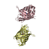
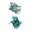
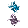
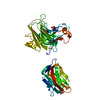
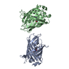
| Title | Crystal structure of the S. cerevisiae YDR533c protein | ||||||
 Components Components | YDR533c protein | ||||||
 Keywords Keywords | structural genomics / unknown function / alpha/beta hydrolase fold / catalytic triad / heat shock protein | ||||||
| Function / homology |  Function and homology information Function and homology informationD-lactate dehydratase / glyoxalase III activity / : / protein folding chaperone / cellular response to nutrient levels / P-body / cytoplasmic stress granule / protein folding / cellular response to oxidative stress / cytoplasm Similarity search - Function | ||||||
| Biological species |  | ||||||
| Method |  X-RAY DIFFRACTION / SYNCHROTRON / MOLECULAR REPLACEMENT / Resolution: 2.35 Å X-RAY DIFFRACTION / SYNCHROTRON / MOLECULAR REPLACEMENT / Resolution: 2.35 Å | ||||||
 Authors Authors | Graille, M. / Leulliot, N. / Quevillon-Cheruel, S. / van Tilbeurgh, H. | ||||||
 Citation Citation | Journal: STRUCTURE / Year: 2004 Title: Crystal structure of the YDR533c S. cerevisiae protein, a class II member of the Hsp31 family Authors: Graille, M. / Quevillon-Cheruel, S. / Leulliot, N. / Zhou, C.Z. / de la Sierra Gallay, I.L. / Jacquamet, L. / Ferrer, J.L. / Liger, D. / Poupon, A. / Janin, J. / van Tilbeurgh, H. | ||||||
| History |
|
- Structure visualization
| Structure viewer | Molecule: MolmilJmol/JSmol |
|---|
- Downloads & links
-Download
| PDBx/mmCIF format | 1qvv.cif.gz | 190.2 KB | Display | PDBx/mmCIF format |
|---|---|---|---|---|
| PDB format | pdb1qvv.ent.gz | 153.9 KB | Display | PDB format |
| PDBx/mmJSON format | 1qvv.json.gz | Tree view | PDBx/mmJSON format | |
| Others |  Other downloads Other downloads |
-Validation report
| Arichive directory | https://data.pdbj.org/pub/pdb/validation_reports/qv/1qvvftp://data.pdbj.org/pub/pdb/validation_reports/qv/1qvv | HTTPS FTP |
|---|
-Related structure data


-Links
 PDBj
PDBj

- Assembly
| Deposited unit | 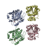
| ||||||||
|---|---|---|---|---|---|---|---|---|---|
| 1 | 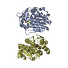
| ||||||||
| 2 | 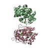
| ||||||||
| Unit cell |
|
-Components
| #1: Protein | Mass: 25777.922 Da / Num. of mol.: 4 Source method: isolated from a genetically manipulated source Source: (gene. exp.) Gene: YDR533c / Plasmid: pET9 / Production host:  #2: Water | ChemComp-HOH / |  Mass: 18.015 Da / Num. of mol.: 325 / Source method: isolated from a natural source / Formula: H2O Mass: 18.015 Da / Num. of mol.: 325 / Source method: isolated from a natural source / Formula: H2OHas protein modification | Y | |
|---|
-Experimental details
-Experiment
| Experiment | Method: X-RAY DIFFRACTION / Number of used crystals: 1 |
|---|
- Sample preparation
| Crystal | Density Matthews: 2.25 Å3/Da / Density % sol: 44.94 % | ||||||||||||||||||||||||
|---|---|---|---|---|---|---|---|---|---|---|---|---|---|---|---|---|---|---|---|---|---|---|---|---|---|
| Crystal grow | Temperature: 293 K / Method: vapor diffusion, hanging drop / pH: 6.4 Details: 26% PEG 4000, 50mM Na/K phosphate buffer, pH 6.4, VAPOR DIFFUSION, HANGING DROP, temperature 293K | ||||||||||||||||||||||||
| Crystal grow | *PLUS Temperature: 18 ℃ / Method: vapor diffusion | ||||||||||||||||||||||||
| Components of the solutions | *PLUS
|
-Data collection
| Diffraction | Mean temperature: 100 K |
|---|---|
| Diffraction source | Source: SYNCHROTRON / Site: ESRF  / Beamline: BM14 / Wavelength: 1.004 Å / Beamline: BM14 / Wavelength: 1.004 Å |
| Detector | Type: MARRESEARCH / Detector: CCD / Date: Nov 29, 2001 |
| Radiation | Monochromator: Si 111 CHANNEL / Protocol: SINGLE WAVELENGTH / Monochromatic (M) / Laue (L): M / Scattering type: x-ray |
| Radiation wavelength | Wavelength: 1.004 Å / Relative weight: 1 |
| Reflection | Resolution: 2.2→30 Å / Num. all: 41388 / Num. obs: 41388 / % possible obs: 84 % / Observed criterion σ(F): 0 / Observed criterion σ(I): 0 / Redundancy: 4.2 % / Biso Wilson estimate: 29.3 Å2 / Rsym value: 0.083 / Net I/σ(I): 7.9 |
| Reflection shell | Resolution: 2.2→2.28 Å / Mean I/σ(I) obs: 1.75 / Rsym value: 0.498 / % possible all: 71.4 |
| Reflection | *PLUS Redundancy: 14.5 % / Num. measured all: 598140 / Rmerge(I) obs: 0.083 |
| Reflection shell | *PLUS % possible obs: 71.4 % / Rmerge(I) obs: 0.498 / Mean I/σ(I) obs: 1.8 |
- Processing
| Software |
| |||||||||||||||||||||||||
|---|---|---|---|---|---|---|---|---|---|---|---|---|---|---|---|---|---|---|---|---|---|---|---|---|---|---|
| Refinement | Method to determine structure: MOLECULAR REPLACEMENT / Resolution: 2.35→29.47 Å / Rfactor Rfree error: 0.007 / Data cutoff high absF: 379163 / Data cutoff low absF: 0 / Isotropic thermal model: Isotropic / Cross valid method: THROUGHOUT / σ(F): 0 / Stereochemistry target values: Engh & Huber
| |||||||||||||||||||||||||
| Solvent computation | Solvent model: FLAT MODEL / Bsol: 36.1282 Å2 / ksol: 0.35398 e/Å3 | |||||||||||||||||||||||||
| Displacement parameters | Biso mean: 31.4 Å2
| |||||||||||||||||||||||||
| Refine analyze |
| |||||||||||||||||||||||||
| Refinement step | Cycle: LAST / Resolution: 2.35→29.47 Å
| |||||||||||||||||||||||||
| Refine LS restraints |
| |||||||||||||||||||||||||
| LS refinement shell | Resolution: 2.35→2.5 Å / Rfactor Rfree error: 0.025 / Total num. of bins used: 6
| |||||||||||||||||||||||||
| Xplor file |
| |||||||||||||||||||||||||
| Refinement | *PLUS Lowest resolution: 30 Å / % reflection Rfree: 5 % | |||||||||||||||||||||||||
| Solvent computation | *PLUS | |||||||||||||||||||||||||
| Displacement parameters | *PLUS | |||||||||||||||||||||||||
| Refine LS restraints | *PLUS
|
