 Movie
Movie Controller


+ Open data

- Basic information
| Entry | Database: PDB / ID: 1qvj | |||||||||
|---|---|---|---|---|---|---|---|---|---|---|
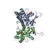
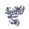
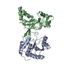
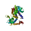
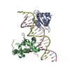
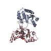
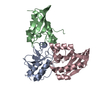
| Title | structure of NUDT9 complexed with ribose-5-phosphate | |||||||||
 Components Components | ADP-ribose pyrophosphatase | |||||||||
 Keywords Keywords | HYDROLASE / NUDIX / ADPRase | |||||||||
| Function / homology |  Function and homology information Function and homology informationADP catabolic process / ADP-sugar pyrophosphatase activity / ADP-ribose diphosphatase / nucleobase-containing small molecule metabolic process / ADP-ribose diphosphatase activity / Phosphate bond hydrolysis by NUDT proteins / cell junction / nuclear membrane / nuclear body / mitochondrial matrix ...ADP catabolic process / ADP-sugar pyrophosphatase activity / ADP-ribose diphosphatase / nucleobase-containing small molecule metabolic process / ADP-ribose diphosphatase activity / Phosphate bond hydrolysis by NUDT proteins / cell junction / nuclear membrane / nuclear body / mitochondrial matrix / mitochondrion / extracellular exosome / cytoplasm / cytosol Similarity search - Function | |||||||||
| Biological species |  Homo sapiens (human) Homo sapiens (human) | |||||||||
| Method |  X-RAY DIFFRACTION / SYNCHROTRON / MOLECULAR REPLACEMENT / Resolution: 1.91 Å X-RAY DIFFRACTION / SYNCHROTRON / MOLECULAR REPLACEMENT / Resolution: 1.91 Å | |||||||||
 Authors Authors | Shen, B.W. / Perraud, A.-L. / Scharenberg, A.S. / Stoddard, B.L. | |||||||||
 Citation Citation | Journal: J.Mol.Biol. / Year: 2003 Title: The crystal structure and mutational analysis of human NUDT9 Authors: Shen, B.W. / Perraud, A.-L. / Scharenberg, A.S. / Stoddard, B.L. #1: Journal: J.Biol.Chem. / Year: 2002Title: NUDT9, a member of the nudix hydrolase family, is an evolutionarily conserved mitochondrial ADP-ribose pyrophosphotase Authors: Perraud, A.L. / Shen, B. / Dunn, C.A. / Rippe, K. / Smith, M.K. / Bessman, M.J. / Stoddard, B.L. / Scharenberg, A.M. | |||||||||
| History |
|
- Structure visualization
| Structure viewer | Molecule: MolmilJmol/JSmol |
|---|
- Downloads & links
-Download
| PDBx/mmCIF format | 1qvj.cif.gz | 79.9 KB | Display | PDBx/mmCIF format |
|---|---|---|---|---|
| PDB format | pdb1qvj.ent.gz | 58.9 KB | Display | PDB format |
| PDBx/mmJSON format | 1qvj.json.gz | Tree view | PDBx/mmJSON format | |
| Others |  Other downloads Other downloads |
-Validation report
| Arichive directory | https://data.pdbj.org/pub/pdb/validation_reports/qv/1qvjftp://data.pdbj.org/pub/pdb/validation_reports/qv/1qvj | HTTPS FTP |
|---|
-Related structure data
| Related structure data |  1q33SC S: Starting model for refinement C: citing same article ( |
|---|---|
| Similar structure data |


-Links
 PDBj
PDBj


- Assembly
| Deposited unit | 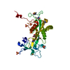
| ||||||||
|---|---|---|---|---|---|---|---|---|---|
| 1 |
| ||||||||
| Unit cell |
|
-Components
-Protein / Sugars , 2 types, 2 molecules A
| #1: Protein | Mass: 33314.457 Da / Num. of mol.: 1 Source method: isolated from a genetically manipulated source Source: (gene. exp.) Homo sapiens (human) / Gene: NUDT9 / Plasmid: pET24b / Production host:  |
|---|---|
| #5: Sugar | ChemComp-RP5 /  Type: D-saccharide, beta linking / Mass: 230.110 Da / Num. of mol.: 1 / Source method: obtained synthetically / Formula: C5H11O8P Type: D-saccharide, beta linking / Mass: 230.110 Da / Num. of mol.: 1 / Source method: obtained synthetically / Formula: C5H11O8P |
-Non-polymers , 4 types, 210 molecules 


| #2: Chemical | ChemComp-SO4 /  Mass: 96.063 Da / Num. of mol.: 5 / Source method: obtained synthetically / Formula: SO4 Mass: 96.063 Da / Num. of mol.: 5 / Source method: obtained synthetically / Formula: SO4#3: Chemical |  Mass: 24.305 Da / Num. of mol.: 2 / Source method: obtained synthetically / Formula: Mg Mass: 24.305 Da / Num. of mol.: 2 / Source method: obtained synthetically / Formula: Mg#4: Chemical | ChemComp-TRS / |  Mass: 122.143 Da / Num. of mol.: 1 / Source method: obtained synthetically / Formula: C4H12NO3 / Comment: pH buffer*YM Mass: 122.143 Da / Num. of mol.: 1 / Source method: obtained synthetically / Formula: C4H12NO3 / Comment: pH buffer*YM#6: Water | ChemComp-HOH / | Mass: 18.015 Da / Num. of mol.: 202 / Source method: isolated from a natural source / Formula: H2O |
|---|
-Details
| Has protein modification | Y |
|---|
-Experimental details
-Experiment
| Experiment | Method: X-RAY DIFFRACTION / Number of used crystals: 1 |
|---|
- Sample preparation
| Crystal | Density Matthews: 3.11 Å3/Da / Density % sol: 60.49 % | |||||||||||||||||||||||||||||||||||||||||||||||||
|---|---|---|---|---|---|---|---|---|---|---|---|---|---|---|---|---|---|---|---|---|---|---|---|---|---|---|---|---|---|---|---|---|---|---|---|---|---|---|---|---|---|---|---|---|---|---|---|---|---|---|
| Crystal grow | Temperature: 298 K / Method: vapor diffusion, hanging drop / pH: 8 Details: Ammonium sulfate, magnisum chloride, trisHCl, pH 8, VAPOR DIFFUSION, HANGING DROP, temperature 298K | |||||||||||||||||||||||||||||||||||||||||||||||||
| Crystal grow | *PLUS pH: 8.5 / Method: vapor diffusion, hanging drop | |||||||||||||||||||||||||||||||||||||||||||||||||
| Components of the solutions | *PLUS
|
-Data collection
| Diffraction | Mean temperature: 170 K |
|---|---|
| Diffraction source | Source: SYNCHROTRON / Site: ALS  / Beamline: 5.0.1 / Wavelength: 1 Å / Beamline: 5.0.1 / Wavelength: 1 Å |
| Detector | Type: ADSC QUANTUM 4 / Detector: CCD / Date: Sep 20, 2002 |
| Radiation | Monochromator: Si 111 CHANNEL / Protocol: SINGLE WAVELENGTH / Monochromatic (M) / Laue (L): M / Scattering type: x-ray |
| Radiation wavelength | Wavelength: 1 Å / Relative weight: 1 |
| Reflection | Resolution: 1.91→41.97 Å / Num. obs: 62416 / % possible obs: 94.8 % / Observed criterion σ(F): 0 / Observed criterion σ(I): 0 / Redundancy: 5.6 % / Biso Wilson estimate: 19.7 Å2 / Rsym value: 0.043 / Net I/σ(I): 35.4 |
| Reflection shell | Resolution: 1.91→1.98 Å / Mean I/σ(I) obs: 4.4 / Num. unique all: 2850 / Rsym value: 0.311 / % possible all: 88.9 |
| Reflection | *PLUS Highest resolution: 1.9 Å / Lowest resolution: 50 Å / Num. obs: 31006 / Num. measured all: 171127 / Rmerge(I) obs: 0.043 |
| Reflection shell | *PLUS % possible obs: 88.9 % / Rmerge(I) obs: 0.311 |
- Processing
| Software |
| ||||||||||||||||||||||||||||||||||||
|---|---|---|---|---|---|---|---|---|---|---|---|---|---|---|---|---|---|---|---|---|---|---|---|---|---|---|---|---|---|---|---|---|---|---|---|---|---|
| Refinement | Method to determine structure: MOLECULAR REPLACEMENT Starting model: 1Q33 Resolution: 1.91→41.97 Å / Rfactor Rfree error: 0.003 / Isotropic thermal model: RESTRAINED / Cross valid method: THROUGHOUT / σ(F): 0 / σ(I): 0 / Stereochemistry target values: Engh & Huber
| ||||||||||||||||||||||||||||||||||||
| Solvent computation | Solvent model: FLAT MODEL / Bsol: 47.5693 Å2 / ksol: 0.383074 e/Å3 | ||||||||||||||||||||||||||||||||||||
| Displacement parameters | Biso mean: 34.5 Å2
| ||||||||||||||||||||||||||||||||||||
| Refine analyze | Luzzati coordinate error free: 0.28 Å / Luzzati sigma a free: 0.25 Å | ||||||||||||||||||||||||||||||||||||
| Refinement step | Cycle: LAST / Resolution: 1.91→41.97 Å
| ||||||||||||||||||||||||||||||||||||
| Refine LS restraints |
| ||||||||||||||||||||||||||||||||||||
| LS refinement shell | Resolution: 1.91→2.03 Å / Rfactor Rfree error: 0.01 / Total num. of bins used: 6
| ||||||||||||||||||||||||||||||||||||
| Xplor file |
| ||||||||||||||||||||||||||||||||||||
| Refinement | *PLUS Highest resolution: 1.9 Å / Lowest resolution: 50 Å / Rfactor Rfree: 0.243 / Rfactor Rwork: 0.215 | ||||||||||||||||||||||||||||||||||||
| Solvent computation | *PLUS | ||||||||||||||||||||||||||||||||||||
| Displacement parameters | *PLUS | ||||||||||||||||||||||||||||||||||||
| Refine LS restraints | *PLUS
|
