 Movie
Movie Controller

[English] 日本語
 Yorodumi
- PDB-1qut: THE SOLUBLE LYTIC TRANSGLYCOSYLASE SLT35 FROM ESCHERICHIA COLI IN... -
+ Open data

- Basic information
| Entry | Database: PDB / ID: 1qut | ||||||
|---|---|---|---|---|---|---|---|
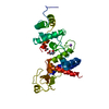
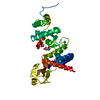
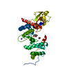
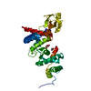
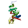
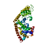
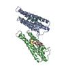
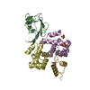
| Title | THE SOLUBLE LYTIC TRANSGLYCOSYLASE SLT35 FROM ESCHERICHIA COLI IN COMPLEX WITH N-ACETYLGLUCOSAMINE | ||||||
 Components Components | LYTIC MUREIN TRANSGLYCOSYLASE B | ||||||
 Keywords Keywords | HYDROLASE / ALPHA-HELICAL PROTEIN WITH A FIVE-STRANDED ANTIPARALLEL BETA-STRAND | ||||||
| Function / homology |  Function and homology information Function and homology informationlytic endotransglycosylase activity / : / peptidoglycan lytic transglycosylase activity / sodium ion binding / peptidoglycan catabolic process / cell outer membrane / cell wall organization / outer membrane-bounded periplasmic space / calcium ion binding Similarity search - Function | ||||||
| Biological species |  | ||||||
| Method |  X-RAY DIFFRACTION / Resolution: 2.44 Å X-RAY DIFFRACTION / Resolution: 2.44 Å | ||||||
 Authors Authors | van Asselt, E.J. / Dijkstra, A.J. / Kalk, K.H. / Takacs, B. / Keck, W. / Dijkstra, B.W. | ||||||
 Citation Citation | Journal: Structure Fold.Des. / Year: 1999 Title: Crystal structure of Escherichia coli lytic transglycosylase Slt35 reveals a lysozyme-like catalytic domain with an EF-hand. Authors: van Asselt, E.J. / Dijkstra, A.J. / Kalk, K.H. / Takacs, B. / Keck, W. / Dijkstra, B.W. #1: Journal: Acta Crystallogr.,Sect.D / Year: 1998Title: Accelerated X-ray structure elucidation of a 36 kDa muramidase/ transglycosylase using wARP Authors: van Asselt, E.J. / Perrakis, A. / Kalk, K.H. / Lamzin, V.S. / Dijkstra, B.W. | ||||||
| History |
|
- Structure visualization
| Structure viewer | Molecule: MolmilJmol/JSmol |
|---|
- Downloads & links
-Download
| PDBx/mmCIF format | 1qut.cif.gz | 80.9 KB | Display | PDBx/mmCIF format |
|---|---|---|---|---|
| PDB format | pdb1qut.ent.gz | 59.1 KB | Display | PDB format |
| PDBx/mmJSON format | 1qut.json.gz | Tree view | PDBx/mmJSON format | |
| Others |  Other downloads Other downloads |
-Validation report
| Arichive directory | https://data.pdbj.org/pub/pdb/validation_reports/qu/1qutftp://data.pdbj.org/pub/pdb/validation_reports/qu/1qut | HTTPS FTP |
|---|
-Related structure data


-Links
 PDBj
PDBj


- Assembly
| Deposited unit | 
| ||||||||||
|---|---|---|---|---|---|---|---|---|---|---|---|
| 1 |
| ||||||||||
| Unit cell |
|
-Components
| #1: Protein | Mass: 36079.660 Da / Num. of mol.: 1 / Fragment: SLT35 / Mutation: L40M, L41V Source method: isolated from a genetically manipulated source Source: (gene. exp.) References: UniProt: P41052, Hydrolases; Glycosylases; Glycosidases, i.e. enzymes that hydrolyse O- and S-glycosyl compounds |
|---|---|
| #2: Sugar | ChemComp-NAG /   Type: D-saccharide, beta linking / Mass: 221.208 Da / Num. of mol.: 1 Type: D-saccharide, beta linking / Mass: 221.208 Da / Num. of mol.: 1Source method: isolated from a genetically manipulated source Formula: C8H15NO6 |
| #3: Chemical | ChemComp-NA /   Mass: 22.990 Da / Num. of mol.: 1 / Source method: obtained synthetically / Formula: Na Mass: 22.990 Da / Num. of mol.: 1 / Source method: obtained synthetically / Formula: Na |
| #4: Water | ChemComp-HOH /  Mass: 18.015 Da / Num. of mol.: 203 / Source method: isolated from a natural source / Formula: H2O Mass: 18.015 Da / Num. of mol.: 203 / Source method: isolated from a natural source / Formula: H2O |
-Experimental details
-Experiment
| Experiment | Method: X-RAY DIFFRACTION / Number of used crystals: 1 |
|---|
- Sample preparation
| Crystal | Density Matthews: 2.7 Å3/Da / Density % sol: 54.5 % | |||||||||||||||
|---|---|---|---|---|---|---|---|---|---|---|---|---|---|---|---|---|
| Crystal grow | Temperature: 295 K / Method: vapor diffusion, hanging drop / pH: 7.8 Details: BICINE-NAOH, PEG 20K, ISOPROPANOL, pH 7.8, VAPOR DIFFUSION, HANGING DROP, temperature 295K | |||||||||||||||
| Crystal | *PLUS Density % sol: 55 % | |||||||||||||||
| Crystal grow | *PLUS Method: unknown / PH range low: 8.5 / PH range high: 7.8 | |||||||||||||||
| Components of the solutions | *PLUS
|
-Data collection
| Diffraction | Mean temperature: 120 K |
|---|---|
| Diffraction source | Source: ROTATING ANODE / Type: ENRAF-NONIUS FR591 / Wavelength: 1.5418 |
| Detector | Type: MAC Science DIP-2000H / Detector: IMAGE PLATE / Date: Oct 28, 1999 |
| Radiation | Protocol: SINGLE WAVELENGTH / Monochromatic (M) / Laue (L): M / Scattering type: x-ray |
| Radiation wavelength | Wavelength: 1.5418 Å / Relative weight: 1 |
| Reflection | Resolution: 2.44→25 Å / Num. obs: 14836 / % possible obs: 98.4 % / Observed criterion σ(I): -3 / Redundancy: 3.3 % / Biso Wilson estimate: 19.5 Å2 / Rmerge(I) obs: 0.104 / Net I/σ(I): 11.1 |
| Reflection shell | Resolution: 2.44→2.48 Å / Redundancy: 2.7 % / Rmerge(I) obs: 0.316 / % possible all: 84 |
| Reflection | *PLUS Num. measured all: 49117 |
- Processing
| Software |
| ||||||||||||||||||||||||||||||||||||||||||||||||||||||||||||||||||||||||||||||||
|---|---|---|---|---|---|---|---|---|---|---|---|---|---|---|---|---|---|---|---|---|---|---|---|---|---|---|---|---|---|---|---|---|---|---|---|---|---|---|---|---|---|---|---|---|---|---|---|---|---|---|---|---|---|---|---|---|---|---|---|---|---|---|---|---|---|---|---|---|---|---|---|---|---|---|---|---|---|---|---|---|---|
| Refinement | Resolution: 2.44→20 Å / Rfactor Rfree error: 0.007 / Data cutoff high absF: 100000 / Data cutoff low absF: 0 / Isotropic thermal model: RESTRAINED / Cross valid method: THROUGHOUT / σ(F): 0 / Stereochemistry target values: ENGH & HUBER
| ||||||||||||||||||||||||||||||||||||||||||||||||||||||||||||||||||||||||||||||||
| Displacement parameters | Biso mean: 21.9 Å2
| ||||||||||||||||||||||||||||||||||||||||||||||||||||||||||||||||||||||||||||||||
| Refine analyze |
| ||||||||||||||||||||||||||||||||||||||||||||||||||||||||||||||||||||||||||||||||
| Refinement step | Cycle: LAST / Resolution: 2.44→20 Å
| ||||||||||||||||||||||||||||||||||||||||||||||||||||||||||||||||||||||||||||||||
| Refine LS restraints |
| ||||||||||||||||||||||||||||||||||||||||||||||||||||||||||||||||||||||||||||||||
| LS refinement shell | Resolution: 2.44→2.55 Å / Rfactor Rfree error: 0.026 / Total num. of bins used: 8
| ||||||||||||||||||||||||||||||||||||||||||||||||||||||||||||||||||||||||||||||||
| Software | *PLUS Name: X-PLOR / Version: 3.843 / Classification: refinement | ||||||||||||||||||||||||||||||||||||||||||||||||||||||||||||||||||||||||||||||||
| Refinement | *PLUS σ(F): 0 / % reflection Rfree: 10.1 % | ||||||||||||||||||||||||||||||||||||||||||||||||||||||||||||||||||||||||||||||||
| Solvent computation | *PLUS | ||||||||||||||||||||||||||||||||||||||||||||||||||||||||||||||||||||||||||||||||
| Displacement parameters | *PLUS Biso mean: 21.9 Å2 | ||||||||||||||||||||||||||||||||||||||||||||||||||||||||||||||||||||||||||||||||
| Refine LS restraints | *PLUS
| ||||||||||||||||||||||||||||||||||||||||||||||||||||||||||||||||||||||||||||||||
| LS refinement shell | *PLUS Rfactor Rfree: 0.348 / % reflection Rfree: 10.7 % / Rfactor Rwork: 0.249 |
