 Movie
Movie Controller

[English] 日本語
 Yorodumi
- PDB-1qr1: POOR BINDING OF A HER-2/NEU EPITOPE (GP2) TO HLA-A2.1 IS DUE TO A... -
+ Open data

- Basic information
| Entry | Database: PDB / ID: 1qr1 | ||||||
|---|---|---|---|---|---|---|---|
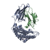
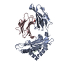
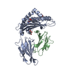
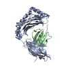
| Title | POOR BINDING OF A HER-2/NEU EPITOPE (GP2) TO HLA-A2.1 IS DUE TO A LACK OF INTERACTIONS IN THE CENTER OF THE PEPTIDE | ||||||
 Components Components |
| ||||||
 Keywords Keywords | IMMUNE SYSTEM / PEPTIDE-BINDING SUPERDOMAIN | ||||||
| Function / homology |  Function and homology information Function and homology informationnegative regulation of immature T cell proliferation in thymus / ERBB3:ERBB2 complex / ERBB2-ERBB4 signaling pathway / immature T cell proliferation in thymus / GRB7 events in ERBB2 signaling / RNA polymerase I core binding / semaphorin receptor complex / Developmental Lineage of Mammary Stem Cells / ErbB-3 class receptor binding / Sema4D induced cell migration and growth-cone collapse ...negative regulation of immature T cell proliferation in thymus / ERBB3:ERBB2 complex / ERBB2-ERBB4 signaling pathway / immature T cell proliferation in thymus / GRB7 events in ERBB2 signaling / RNA polymerase I core binding / semaphorin receptor complex / Developmental Lineage of Mammary Stem Cells / ErbB-3 class receptor binding / Sema4D induced cell migration and growth-cone collapse / motor neuron axon guidance / regulation of microtubule-based process / positive regulation of memory T cell activation / T cell mediated cytotoxicity directed against tumor cell target / positive regulation of CD8-positive, alpha-beta T cell activation / CD8-positive, alpha-beta T cell activation / positive regulation of CD8-positive, alpha-beta T cell proliferation / PLCG1 events in ERBB2 signaling / enzyme-linked receptor protein signaling pathway / ERBB2 Activates PTK6 Signaling / antigen processing and presentation of endogenous peptide antigen via MHC class I via ER pathway, TAP-dependent / TAP complex binding / ERBB2-EGFR signaling pathway / antigen processing and presentation of exogenous peptide antigen via MHC class I / Golgi medial cisterna / ERBB2-ERBB3 signaling pathway / neurotransmitter receptor localization to postsynaptic specialization membrane / Drug-mediated inhibition of ERBB2 signaling / Resistance of ERBB2 KD mutants to trastuzumab / Resistance of ERBB2 KD mutants to sapitinib / Resistance of ERBB2 KD mutants to tesevatinib / Resistance of ERBB2 KD mutants to neratinib / Resistance of ERBB2 KD mutants to osimertinib / Resistance of ERBB2 KD mutants to afatinib / Resistance of ERBB2 KD mutants to AEE788 / Resistance of ERBB2 KD mutants to lapatinib / Drug resistance in ERBB2 TMD/JMD mutants / positive regulation of MAP kinase activity / neuromuscular junction development / positive regulation of Rho protein signal transduction / positive regulation of transcription by RNA polymerase I / CD8 receptor binding / ERBB2 Regulates Cell Motility / Developmental Lineage of Mammary Gland Myoepithelial Cells / oligodendrocyte differentiation / protection from natural killer cell mediated cytotoxicity / semaphorin-plexin signaling pathway / beta-2-microglobulin binding / endoplasmic reticulum exit site / PI3K events in ERBB2 signaling / TAP binding / regulation of angiogenesis / positive regulation of protein targeting to membrane / detection of bacterium / antigen processing and presentation of endogenous peptide antigen via MHC class Ib / antigen processing and presentation of endogenous peptide antigen via MHC class I via ER pathway, TAP-independent / regulation of ERK1 and ERK2 cascade / Schwann cell development / coreceptor activity / T cell receptor binding / Signaling by ERBB2 / TFAP2 (AP-2) family regulates transcription of growth factors and their receptors / peptidyl-tyrosine phosphorylation / myelination / transmembrane receptor protein tyrosine kinase activity / GRB2 events in ERBB2 signaling / positive regulation of cell adhesion / SHC1 events in ERBB2 signaling / cell surface receptor protein tyrosine kinase signaling pathway / basal plasma membrane / early endosome lumen / cellular response to epidermal growth factor stimulus / Nef mediated downregulation of MHC class I complex cell surface expression / DAP12 interactions / Constitutive Signaling by Overexpressed ERBB2 / positive regulation of epithelial cell proliferation / Downregulation of ERBB2:ERBB3 signaling / Endosomal/Vacuolar pathway / T cell mediated cytotoxicity / positive regulation of translation / Antigen Presentation: Folding, assembly and peptide loading of class I MHC / lumenal side of endoplasmic reticulum membrane / regulation of iron ion transport / cellular response to iron(III) ion / neuromuscular junction / negative regulation of iron ion transport / negative regulation of forebrain neuron differentiation / antigen processing and presentation of exogenous protein antigen via MHC class Ib, TAP-dependent / wound healing / phosphatidylinositol 3-kinase/protein kinase B signal transduction / peptide antigen assembly with MHC class I protein complex / ER to Golgi transport vesicle membrane / regulation of erythrocyte differentiation / response to molecule of bacterial origin / HFE-transferrin receptor complex / MHC class I peptide loading complex / transferrin transport / Signaling by ERBB2 TMD/JMD mutants / cellular response to iron ion / negative regulation of receptor-mediated endocytosis Similarity search - Function | ||||||
| Biological species |  Homo sapiens (human) Homo sapiens (human) | ||||||
| Method |  X-RAY DIFFRACTION / SYNCHROTRON / Resolution: 2.4 Å X-RAY DIFFRACTION / SYNCHROTRON / Resolution: 2.4 Å | ||||||
 Authors Authors | Kuhns, J.J. / Batalia, M.A. / Yan, S. / Collins, E.J. | ||||||
 Citation Citation | Journal: J.Biol.Chem. / Year: 1999 Title: Poor binding of a HER-2/neu epitope (GP2) to HLA-A2.1 is due to a lack of interactions with the center of the peptide. Authors: Kuhns, J.J. / Batalia, M.A. / Yan, S. / Collins, E.J. | ||||||
| History |
|
- Structure visualization
| Structure viewer | Molecule: MolmilJmol/JSmol |
|---|
- Downloads & links
-Download
| PDBx/mmCIF format | 1qr1.cif.gz | 164.1 KB | Display | PDBx/mmCIF format |
|---|---|---|---|---|
| PDB format | pdb1qr1.ent.gz | 130.4 KB | Display | PDB format |
| PDBx/mmJSON format | 1qr1.json.gz | Tree view | PDBx/mmJSON format | |
| Others |  Other downloads Other downloads |
-Validation report
| Arichive directory | https://data.pdbj.org/pub/pdb/validation_reports/qr/1qr1ftp://data.pdbj.org/pub/pdb/validation_reports/qr/1qr1 | HTTPS FTP |
|---|
-Related structure data
| Related structure data | |
|---|---|
| Similar structure data |


-Links
 PDBj
PDBj


- Assembly
| Deposited unit | 
| ||||||||||
|---|---|---|---|---|---|---|---|---|---|---|---|
| 1 | 
| ||||||||||
| 2 | 
| ||||||||||
| Unit cell |
|
-Components
| #1: Protein | Mass: 31854.203 Da / Num. of mol.: 2 / Fragment: RESIDUES 1-275 OF EXTRACELLULAR PORTION Source method: isolated from a genetically manipulated source Source: (gene. exp.) Homo sapiens (human) / Plasmid details: T7 promoter system / Plasmid: pLM1 / Production host:  #2: Protein | Mass: 11879.356 Da / Num. of mol.: 2 Mutation: B2M HAS AN ADDED METHIONINE FOR BACTERIAL EXPRESSION Source method: isolated from a genetically manipulated source Source: (gene. exp.) Homo sapiens (human) / Plasmid details: T7 promoter system / Plasmid: pLM1 / Production host: #3: Protein/peptide | Mass: 884.115 Da / Num. of mol.: 2 / Source method: obtained synthetically / Details: synthetic peptide by FMOC chemistry / References: UniProt: P04626 #4: Water | ChemComp-HOH / |  Mass: 18.015 Da / Num. of mol.: 103 / Source method: isolated from a natural source / Formula: H2O Mass: 18.015 Da / Num. of mol.: 103 / Source method: isolated from a natural source / Formula: H2OHas protein modification | Y | |
|---|
-Experimental details
-Experiment
| Experiment | Method: X-RAY DIFFRACTION / Number of used crystals: 1 |
|---|
- Sample preparation
| Crystal | Density Matthews: 2.55 Å3/Da / Density % sol: 51.71 % | ||||||||||||||||||||||||||||||
|---|---|---|---|---|---|---|---|---|---|---|---|---|---|---|---|---|---|---|---|---|---|---|---|---|---|---|---|---|---|---|---|
| Crystal grow | Temperature: 295 K / Method: vapor diffusion, hanging drop / pH: 6.5 Details: 14% PEG 6000, 25 mM MES, pH 6.5, VAPOR DIFFUSION, HANGING DROP, temperature 22K | ||||||||||||||||||||||||||||||
| Crystal grow | *PLUS Details: Zhao, R., (1999) J.Exp.Med., 189, 359. | ||||||||||||||||||||||||||||||
| Components of the solutions | *PLUS
|
-Data collection
| Diffraction | Mean temperature: 100 K |
|---|---|
| Diffraction source | Source: SYNCHROTRON / Site: NSLS  / Beamline: X12B / Wavelength: 0.91 / Beamline: X12B / Wavelength: 0.91 |
| Detector | Type: ADSC QUANTUM 4 / Detector: CCD / Date: Jun 10, 1998 |
| Radiation | Protocol: SINGLE WAVELENGTH / Monochromatic (M) / Laue (L): M / Scattering type: x-ray |
| Radiation wavelength | Wavelength: 0.91 Å / Relative weight: 1 |
| Reflection | Resolution: 2.5→30 Å / Num. all: 34412 / % possible obs: 98.2 % / Observed criterion σ(F): 0 / Observed criterion σ(I): -3 / Redundancy: 2 % / Biso Wilson estimate: 17.4 Å2 / Rmerge(I) obs: 0.093 / Net I/σ(I): 7.8 |
| Reflection shell | Resolution: 2.4→2.44 Å / Redundancy: 2 % / Rmerge(I) obs: 0.233 / Num. unique all: 1900 / % possible all: 97.6 |
| Reflection | *PLUS Num. obs: 34962 / Num. measured all: 66839 |
| Reflection shell | *PLUS Highest resolution: 2.4 Å / % possible obs: 97.6 % / Mean I/σ(I) obs: 3.46 |
- Processing
| Software |
| |||||||||||||||||||||||||
|---|---|---|---|---|---|---|---|---|---|---|---|---|---|---|---|---|---|---|---|---|---|---|---|---|---|---|
| Refinement | Resolution: 2.4→30 Å / Rfactor Rfree error: 0.008 / Data cutoff high absF: 997316.97 / Data cutoff low absF: 0 / Isotropic thermal model: RESTRAINED / Cross valid method: THROUGHOUT / σ(F): 0 / σ(I): -3 / Stereochemistry target values: Engh & Huber
| |||||||||||||||||||||||||
| Solvent computation | Solvent model: FLAT MODEL / Bsol: 19.99 Å2 / ksol: 0.343 e/Å3 | |||||||||||||||||||||||||
| Displacement parameters | Biso mean: 17.5 Å2
| |||||||||||||||||||||||||
| Refine analyze |
| |||||||||||||||||||||||||
| Refinement step | Cycle: LAST / Resolution: 2.4→30 Å
| |||||||||||||||||||||||||
| Refine LS restraints |
| |||||||||||||||||||||||||
| LS refinement shell | Resolution: 2.4→2.55 Å / Rfactor Rfree error: 0.026 / Total num. of bins used: 6
| |||||||||||||||||||||||||
| Xplor file |
| |||||||||||||||||||||||||
| Software | *PLUS Name: CNS / Version: 0.5 / Classification: refinement | |||||||||||||||||||||||||
| Refinement | *PLUS Lowest resolution: 30 Å / σ(F): 0 / % reflection Rfree: 5.1 % / Rfactor obs: 0.242 | |||||||||||||||||||||||||
| Solvent computation | *PLUS | |||||||||||||||||||||||||
| Displacement parameters | *PLUS Biso mean: 17.5 Å2 | |||||||||||||||||||||||||
| Refine LS restraints | *PLUS
| |||||||||||||||||||||||||
| LS refinement shell | *PLUS Rfactor Rfree: 0.285 / % reflection Rfree: 5 % / Rfactor Rwork: 0.362 |
