 Movie
Movie Controller

[English] 日本語
 Yorodumi
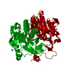
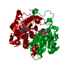
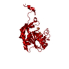
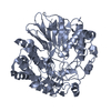
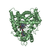
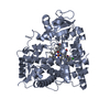
- PDB-1qcc: CRYSTAL STRUCTURES OF ADENINE PHOSPHORIBOSYLTRANSFERASE FROM LEIS... -
+ Open data

- Basic information
| Entry | Database: PDB / ID: 1qcc | ||||||
|---|---|---|---|---|---|---|---|
| Title | CRYSTAL STRUCTURES OF ADENINE PHOSPHORIBOSYLTRANSFERASE FROM LEISHMANIA DONOVANI | ||||||
 Components Components | ADENINE PHOSPHORIBOSYLTRANSFERASE | ||||||
 Keywords Keywords | TRANSFERASE / DINUCLEOTIDE BINDING FOLD | ||||||
| Function / homology |  Function and homology information Function and homology informationadenine phosphoribosyltransferase / adenine phosphoribosyltransferase activity / purine ribonucleoside salvage / nucleotide binding / metal ion binding / cytoplasm Similarity search - Function | ||||||
| Biological species |  Leishmania donovani (eukaryote) Leishmania donovani (eukaryote) | ||||||
| Method |  X-RAY DIFFRACTION / Resolution: 1.98 Å X-RAY DIFFRACTION / Resolution: 1.98 Å | ||||||
 Authors Authors | Phillips, C.L. / Ullman, B. / Brennan, R.G. / Hill, C.P. | ||||||
 Citation Citation | Journal: EMBO J. / Year: 1999 Title: Crystal structures of adenine phosphoribosyltransferase from Leishmania donovani. Authors: Phillips, C.L. / Ullman, B. / Brennan, R.G. / Hill, C.P. | ||||||
| History |
|
- Structure visualization
| Structure viewer | Molecule: MolmilJmol/JSmol |
|---|
- Downloads & links
-Download
| PDBx/mmCIF format | 1qcc.cif.gz | 62.2 KB | Display | PDBx/mmCIF format |
|---|---|---|---|---|
| PDB format | pdb1qcc.ent.gz | 45.5 KB | Display | PDB format |
| PDBx/mmJSON format | 1qcc.json.gz | Tree view | PDBx/mmJSON format | |
| Others |  Other downloads Other downloads |
-Validation report
| Arichive directory | https://data.pdbj.org/pub/pdb/validation_reports/qc/1qccftp://data.pdbj.org/pub/pdb/validation_reports/qc/1qcc | HTTPS FTP |
|---|
-Related structure data


-Links
 PDBj
PDBj
- Assembly
| Deposited unit | 
| ||||||||
|---|---|---|---|---|---|---|---|---|---|
| 1 | 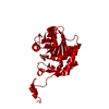
| ||||||||
| Unit cell |
|
-Components
| #1: Protein | Mass: 26096.920 Da / Num. of mol.: 1 Source method: isolated from a genetically manipulated source Source: (gene. exp.) Leishmania donovani (eukaryote) / Production host:  |
|---|---|
| #2: Chemical | ChemComp-CIT /   Mass: 192.124 Da / Num. of mol.: 1 / Source method: obtained synthetically / Formula: C6H8O7 Mass: 192.124 Da / Num. of mol.: 1 / Source method: obtained synthetically / Formula: C6H8O7 |
| #3: Water | ChemComp-HOH /  Mass: 18.015 Da / Num. of mol.: 215 / Source method: isolated from a natural source / Formula: H2O Mass: 18.015 Da / Num. of mol.: 215 / Source method: isolated from a natural source / Formula: H2O |
-Experimental details
-Experiment
| Experiment | Method: X-RAY DIFFRACTION / Number of used crystals: 1 |
|---|
- Sample preparation
| Crystal | Density Matthews: 2.66 Å3/Da / Density % sol: 53.73 % | ||||||||||||||||||||||||||||||||||||||||||||||||||||||
|---|---|---|---|---|---|---|---|---|---|---|---|---|---|---|---|---|---|---|---|---|---|---|---|---|---|---|---|---|---|---|---|---|---|---|---|---|---|---|---|---|---|---|---|---|---|---|---|---|---|---|---|---|---|---|---|
| Crystal grow | Temperature: 277 K / Method: vapor diffusion, hanging drop / pH: 5 Details: REG 5000 MME, AMMONIUM ACETATE, SODIUM CITRATE, MAGNESIUM CHLORIDE, pH 5.0, VAPOR DIFFUSION, HANGING DROP, temperature 277.0K | ||||||||||||||||||||||||||||||||||||||||||||||||||||||
| Crystal grow | *PLUS Temperature: 4 ℃ / pH: 6 | ||||||||||||||||||||||||||||||||||||||||||||||||||||||
| Components of the solutions | *PLUS
|
-Data collection
| Diffraction | Mean temperature: 100 K |
|---|---|
| Diffraction source | Source: ROTATING ANODE / Type: RIGAKU RU200 / Wavelength: 1.5418 |
| Detector | Type: RIGAKU RAXIS IV / Detector: IMAGE PLATE / Date: Feb 1, 1997 |
| Radiation | Protocol: SINGLE WAVELENGTH / Monochromatic (M) / Laue (L): M / Scattering type: x-ray |
| Radiation wavelength | Wavelength: 1.5418 Å / Relative weight: 1 |
| Reflection | Biso Wilson estimate: 22.3 Å2 |
| Reflection | *PLUS Highest resolution: 1.98 Å / Lowest resolution: 19.7 Å / Num. obs: 19055 / % possible obs: 91.5 % / Num. measured all: 349744 / Rmerge(I) obs: 0.051 |
| Reflection shell | *PLUS % possible obs: 61.3 % / Rmerge(I) obs: 0.124 / Mean I/σ(I) obs: 10.2 |
- Processing
| Software |
| ||||||||||||||||||||||||||||||||||||||||||||||||||||||||||||
|---|---|---|---|---|---|---|---|---|---|---|---|---|---|---|---|---|---|---|---|---|---|---|---|---|---|---|---|---|---|---|---|---|---|---|---|---|---|---|---|---|---|---|---|---|---|---|---|---|---|---|---|---|---|---|---|---|---|---|---|---|---|
| Refinement | Resolution: 1.98→19.7 Å / Rfactor Rfree error: 0.006 / Data cutoff high absF: 10000000 / Data cutoff low absF: 0 / Isotropic thermal model: RESTRAINED / Cross valid method: THROUGHOUT / σ(F): 0
| ||||||||||||||||||||||||||||||||||||||||||||||||||||||||||||
| Displacement parameters | Biso mean: 23.8 Å2
| ||||||||||||||||||||||||||||||||||||||||||||||||||||||||||||
| Refine analyze |
| ||||||||||||||||||||||||||||||||||||||||||||||||||||||||||||
| Refinement step | Cycle: LAST / Resolution: 1.98→19.7 Å
| ||||||||||||||||||||||||||||||||||||||||||||||||||||||||||||
| Refine LS restraints |
| ||||||||||||||||||||||||||||||||||||||||||||||||||||||||||||
| LS refinement shell | Resolution: 1.98→2.1 Å / Rfactor Rfree error: 0.02 / Total num. of bins used: 6
| ||||||||||||||||||||||||||||||||||||||||||||||||||||||||||||
| Software | *PLUS Name: X-PLOR / Version: 3.843 / Classification: refinement | ||||||||||||||||||||||||||||||||||||||||||||||||||||||||||||
| Refine LS restraints | *PLUS
|
