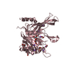
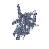
Mass: 12601.585 Da / Num. of mol.: 2 / Fragment: C-terminal domain (ThKaiA180C) Source method: isolated from a genetically manipulated source Source: (gene. exp.) Thermosynechococcus elongatus (bacteria) Strain: BP-1 / Gene: KaiA / Plasmid: pET-32a / Species (production host): Escherichia coli / Production host: Escherichia coli BL21(DE3) (bacteria) / Strain (production host): BL21(DE3) / References: UniProt: Q8RR35, UniProt: Q79V62*PLUS
Has protein modification
Y
-
Experimental details
-
Experiment
Experiment
Method: SOLUTION NMR
NMR experiment
Conditions-ID
Experiment-ID
Solution-ID
Type
1
1
1
3D 15N-separated NOESY
1
2
3
4D 13C-separated NOESY
1
3
4
3D 13C-edited, 12C filtered NOESY
1
4
3
HACAHB
1
5
1
HNHB
1
6
3
BRCTCO/CN
1
7
2
CBCA(CO)NH
1
8
2
CBCANH
NMR details
Text: This is the oxidized form of the protein. A disulfide bond connects residue C96 of each monomeric unit.
-
Sample preparation
Details
Solution-ID
Contents
Solvent system
1
1.3 mM ThKaiA180C U-15N, 20 mM NaCl, 20 mM phosphate buffer, 95% H2O, 5% D2O
95% H2O/5% D2O
2
1.3 mM ThKaiA180C U-15N,U-13C, 20 mM NaCl, 20 mM phosphate buffer, 95% H2O, 5% D2O
95% H2O/5% D2O
3
1.3 mM ThKaiA180C U-15N,U-13C, 20 mM NaCl, 20 mM phosphate buffer, 100% D2O
100% D2O
4
1.0 mM ThKaiA180C U-15N,U-13C, 1.0 mM ThKaiA180C NA-N,Na-C, 20 mM NaCl, 20 mM phosphate buffer, 100% D2O
100% D2O
Sample conditions
Ionic strength: 20 mM NaCl, 20 mM NaPi / pH: 7 / Pressure: ambient / Temperature: 323 K
-
NMR measurement
Radiation
Protocol: SINGLE WAVELENGTH / Monochromatic (M) / Laue (L): M / Scattering type: x-ray
Radiation wavelength
Relative weight: 1
NMR spectrometer
Type
Manufacturer
Model
Field strength (MHz)
Spectrometer-ID
Varian INOVA
Varian
INOVA
600
1
Varian INOVA
Varian
INOVA
500
2
-
Processing
NMR software
Name
Version
Developer
Classification
NMRPipe
2.1Rev2002.044.17.08
Delaglio
processing
PIPP
4.2.6
Garrett
dataanalysis
XPLOR-NIH
2.9.1
Clore
structuresolution
VNMR
6.1Rev. C
VarianAssoc.
collection
XPLOR-NIH
2.9.1
Clore
refinement
Refinement
Method: Distance geometry, Simulating annealing, Radius-of-gyration, Carbon chemical shift, conformational database potential refinement Software ordinal: 1 Details: The structure is based on 2207 restraints per monomeric unit of which 1740 are NOE restraints.
NMR representative
Selection criteria: closest to the average
NMR ensemble
Conformer selection criteria: structures with the least restraint violations Conformers calculated total number: 50 / Conformers submitted total number: 25
+
About Yorodumi
-
News
-
Feb 9, 2022. New format data for meta-information of EMDB entries
New format data for meta-information of EMDB entries
Version 3 of the EMDB header file is now the official format.
The previous official version 1.9 will be removed from the archive.
In the structure databanks used in Yorodumi, some data are registered as the other names, "COVID-19 virus" and "2019-nCoV". Here are the details of the virus and the list of structure data.
Jan 31, 2019. EMDB accession codes are about to change! (news from PDBe EMDB page)
EMDB accession codes are about to change! (news from PDBe EMDB page)
The allocation of 4 digits for EMDB accession codes will soon come to an end. Whilst these codes will remain in use, new EMDB accession codes will include an additional digit and will expand incrementally as the available range of codes is exhausted. The current 4-digit format prefixed with “EMD-” (i.e. EMD-XXXX) will advance to a 5-digit format (i.e. EMD-XXXXX), and so on. It is currently estimated that the 4-digit codes will be depleted around Spring 2019, at which point the 5-digit format will come into force.
The EM Navigator/Yorodumi systems omit the EMD- prefix.
Related info.:Q: What is EMD? / ID/Accession-code notation in Yorodumi/EM Navigator
Yorodumi is a browser for structure data from EMDB, PDB, SASBDB, etc.
This page is also the successor to EM Navigator detail page, and also detail information page/front-end page for Omokage search.
The word "yorodu" (or yorozu) is an old Japanese word meaning "ten thousand". "mi" (miru) is to see.
Related info.:EMDB / PDB / SASBDB / Comparison of 3 databanks / Yorodumi Search / Aug 31, 2016. New EM Navigator & Yorodumi / Yorodumi Papers / Jmol/JSmol / Function and homology information / Changes in new EM Navigator and Yorodumi
 Movie
Movie Controller

 Yorodumi
 Open data
 Basic information
 Components
 Keywords
 Function and homology information
 Thermosynechococcus elongatus (bacteria)
 Authors
 Citation
 Structure visualization
 Downloads & links
 Other downloads
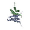


 PDBj
PDBj

 Assembly
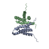
 Sample preparation
 Processing
 NMRPipe