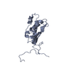
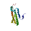
- PDB-1q48: Solution NMR Structure of The Haemophilus Influenzae Iron-Sulfur ... -
+
Open data
ID or keywords:
Loading...
-
Basic information
Entry
Database: PDB / ID: 1q48
Title
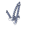
Solution NMR Structure of The Haemophilus Influenzae Iron-Sulfur Cluster Assembly Protein U (IscU) with Zinc Bound at the Active Site. Northeast Structural Genomics Consortium Target IR24. This protein is not apo, it is a model without zinc binding constraints.
COMPOUND THIS PROTEIN HAS STOICHIOMETRIC ZINC BOUND. THIS PDB ENTRY IS A MODEL CALCULATED WITHOUT ...COMPOUND THIS PROTEIN HAS STOICHIOMETRIC ZINC BOUND. THIS PDB ENTRY IS A MODEL CALCULATED WITHOUT THE ZINC-BINDING CONSTRIANTS. PDB ENTRY 1R9P IS THE SAME PROTEIN MODELED WITH ZINC-BINDING CONSTRAINTS OBTAINED BY INDIRECT METHODS.
Method: SIMULATED ANNEALING, TORSION ANGLE DYNAMICS, AUTOMATED ANALYSIS OF NOESY DATA, 3D STRUCTURES Software ordinal: 1 Details: THE STRUCTURES ARE BASED ON A TOTAL OF 923 RESTRAINTS. SUMMARY OF EXPERIMENTAL CONSTRAINTS: DISTANCE CONSTRAINTS: TOTAL = 790; INTRA-RESIDUE [I=J] = 12; SEQUENTIAL [(I-J)=1] = 260; MEDIUM ...Details: THE STRUCTURES ARE BASED ON A TOTAL OF 923 RESTRAINTS. SUMMARY OF EXPERIMENTAL CONSTRAINTS: DISTANCE CONSTRAINTS: TOTAL = 790; INTRA-RESIDUE [I=J] = 12; SEQUENTIAL [(I-J)=1] = 260; MEDIUM RANGE [1<(I-J)<5] = 185; LONG RANGE [(I-J)>=5] = 255; HYDROGEN BOND CONSTRAINTS = 58 (2 PER H-BOND); NUMBER OF DISTANCE CONSTRAINTS PER RESIDUE = 8.0; DIHEDRAL-ANGLE CONSTRAINTS = 133 (66 PHI, 67 PSI); TOTAL NUMBER OF CONSTRAINTS PER RESIDUE = 9.4 (RESIDES 26-123); NUMBER OF LONG RANGE CONSTRAINTS PER RESIDUE = 2.6; NUMBER OF STRUCTURES COMPUTED = 25; NUMBER OF STRUCTURES USED = 20. AVERAGE DISTANCE VIOLATIONS >0.0001 ANG = 24.3; AVERAGE R.M.S. DISTANCE VIOLATION = 0.003 ANG; MAXIMUM NUMBER OF DISTANCE VIOLATIONS 31. AVERAGE DIHEDRAL ANGLE VIOLATIONS: >0.0001 DEG = 1.0; MAX NUMBER OF DIHEDRAL ANGLE VIOLATIONS = 4; AVERAGE R.M.S. ANGLE VIOLATION = 0.01 DEG. RMSD VALUES: BACKBONE ATOMS (N,C,C',O) = 0.80 ANG; ALL HEAVY ATOMS = 1.17 ANG; PROCHECK: MOST FAVORED REGIONS = 77%; ADDITIONAL ALLOWED REGIONS = 20%; GENEROUSLY ALLOWED REGIONS = 3%; DISALLOWED REGIONS = 0%.
NMR representative
Selection criteria: lowest energy
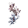
NMR ensemble
Conformer selection criteria: structures with the lowest energy Conformers calculated total number: 25 / Conformers submitted total number: 20
+
About Yorodumi
-
News
-
Feb 9, 2022. New format data for meta-information of EMDB entries
New format data for meta-information of EMDB entries
Version 3 of the EMDB header file is now the official format.
The previous official version 1.9 will be removed from the archive.
In the structure databanks used in Yorodumi, some data are registered as the other names, "COVID-19 virus" and "2019-nCoV". Here are the details of the virus and the list of structure data.
Jan 31, 2019. EMDB accession codes are about to change! (news from PDBe EMDB page)
EMDB accession codes are about to change! (news from PDBe EMDB page)
The allocation of 4 digits for EMDB accession codes will soon come to an end. Whilst these codes will remain in use, new EMDB accession codes will include an additional digit and will expand incrementally as the available range of codes is exhausted. The current 4-digit format prefixed with “EMD-” (i.e. EMD-XXXX) will advance to a 5-digit format (i.e. EMD-XXXXX), and so on. It is currently estimated that the 4-digit codes will be depleted around Spring 2019, at which point the 5-digit format will come into force.
The EM Navigator/Yorodumi systems omit the EMD- prefix.
Related info.:Q: What is EMD? / ID/Accession-code notation in Yorodumi/EM Navigator
Yorodumi is a browser for structure data from EMDB, PDB, SASBDB, etc.
This page is also the successor to EM Navigator detail page, and also detail information page/front-end page for Omokage search.
The word "yorodu" (or yorozu) is an old Japanese word meaning "ten thousand". "mi" (miru) is to see.
Related info.:EMDB / PDB / SASBDB / Comparison of 3 databanks / Yorodumi Search / Aug 31, 2016. New EM Navigator & Yorodumi / Yorodumi Papers / Jmol/JSmol / Function and homology information / Changes in new EM Navigator and Yorodumi
 Movie
Movie Controller

 Yorodumi
 Open data
 Basic information
 Components
 Keywords
 Function and homology information
 Haemophilus influenzae (bacteria)
 Authors
 Citation
 Structure visualization
 Downloads & links
 Other downloads
 PDBj
PDBj Assembly
 HSQC
HSQC Sample preparation
 Processing