 Movie
Movie Controller

[English] 日本語
 Yorodumi
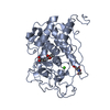
- PDB-1pxz: 1.7 Angstrom Crystal Structure of jun a 1, the major allergen fro... -
+ Open data

- Basic information
| Entry | Database: PDB / ID: 1pxz | ||||||
|---|---|---|---|---|---|---|---|
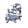
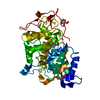
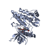
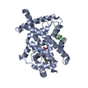
| Title | 1.7 Angstrom Crystal Structure of jun a 1, the major allergen from cedar pollen | ||||||
 Components Components | Major pollen allergen Jun a 1 | ||||||
 Keywords Keywords | ALLERGEN / parallel beta-helix / cedar pollen | ||||||
| Function / homology |  Function and homology information Function and homology informationpectate lyase / pectate lyase activity / pectin catabolic process / metal ion binding Similarity search - Function | ||||||
| Biological species |  Juniperus ashei (Ozark white cedar) Juniperus ashei (Ozark white cedar) | ||||||
| Method |  X-RAY DIFFRACTION / SYNCHROTRON / MIR / Resolution: 1.7 Å X-RAY DIFFRACTION / SYNCHROTRON / MIR / Resolution: 1.7 Å | ||||||
 Authors Authors | Czerwinski, E.W. / White, M.A. / Midoro-Horiuti, T. / Brooks, E.G. / Goldblum, R.M. | ||||||
 Citation Citation | Journal: J.Biol.Chem. / Year: 2005 Title: Crystal structure of Jun a 1, the major cedar pollen allergen from Juniperus ashei, reveals a parallel beta-helical core. Authors: Czerwinski, E.W. / Midoro-Horiuti, T. / White, M.A. / Brooks, E.G. / Goldblum, R.M. #1: Journal: Acta Crystallogr.,Sect.D / Year: 2003Title: Crystallization and preliminary X-ray diffraction analysis of Jun a 1, the major allergen isolated from pollen of the mountain cedar Juniperus ashei Authors: Liu, D. / Midoro-Horiuti, T. / White, M.A. / Brooks, E.G. / Goldblum, R.M. / Czerwinski, E.W. | ||||||
| History |
|
- Structure visualization
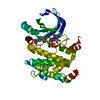
| Structure viewer | Molecule: MolmilJmol/JSmol |
|---|
- Downloads & links
-Download
| PDBx/mmCIF format | 1pxz.cif.gz | 157 KB | Display | PDBx/mmCIF format |
|---|---|---|---|---|
| PDB format | pdb1pxz.ent.gz | 123.7 KB | Display | PDB format |
| PDBx/mmJSON format | 1pxz.json.gz | Tree view | PDBx/mmJSON format | |
| Others |  Other downloads Other downloads |
-Validation report
| Arichive directory | https://data.pdbj.org/pub/pdb/validation_reports/px/1pxzftp://data.pdbj.org/pub/pdb/validation_reports/px/1pxz | HTTPS FTP |
|---|
-Related structure data
| Similar structure data |
|---|


-Links
 PDBj
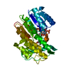
PDBj- Assembly
| Deposited unit |
| ||||||||
|---|---|---|---|---|---|---|---|---|---|
| 1 |
| ||||||||
| 2 |
| ||||||||
| Unit cell |
|
-Components
| #1: Protein | Mass: 37680.867 Da / Num. of mol.: 2 / Source method: isolated from a natural source / Details: pollen / Source: (natural) Juniperus ashei (Ozark white cedar) / References: UniProt: P81294#2: Water | ChemComp-HOH / |  Mass: 18.015 Da / Num. of mol.: 701 / Source method: isolated from a natural source / Formula: H2O Mass: 18.015 Da / Num. of mol.: 701 / Source method: isolated from a natural source / Formula: H2OHas protein modification | Y | |
|---|
-Experimental details
-Experiment
| Experiment | Method: X-RAY DIFFRACTION / Number of used crystals: 1 |
|---|
- Sample preparation
| Crystal | Density Matthews: 2.56 Å3/Da / Density % sol: 58.91 % |
|---|---|
| Crystal grow | Temperature: 277 K / Method: vapor diffusion, hanging drop / pH: 5.5 Details: PEG 4000, ammonium acetate, sodium acetate, pH 5.5, VAPOR DIFFUSION, HANGING DROP, temperature 277K |
-Data collection
| Diffraction | Mean temperature: 100 K |
|---|---|
| Diffraction source | Source: SYNCHROTRON / Site: APS  / Beamline: 14-BM-C / Wavelength: 0.9 / Wavelength: 0.9 Å / Beamline: 14-BM-C / Wavelength: 0.9 / Wavelength: 0.9 Å |
| Detector | Type: ADSC QUANTUM 4 / Detector: CCD / Date: Nov 13, 2002 |
| Radiation | Monochromator: SI / Protocol: SINGLE WAVELENGTH / Monochromatic (M) / Laue (L): M / Scattering type: x-ray |
| Radiation wavelength | Wavelength: 0.9 Å / Relative weight: 1 |
| Reflection | Resolution: 1.7→100 Å / Num. obs: 89001 / % possible obs: 91.3 % / Observed criterion σ(F): 0 / Observed criterion σ(I): 0 / Redundancy: 3.2 % / Biso Wilson estimate: 21.3 Å2 / Rsym value: 0.05 / Net I/σ(I): 14.5 |
| Reflection shell | Resolution: 1.7→1.74 Å / Redundancy: 2 % / Mean I/σ(I) obs: 4.1 / Num. unique all: 3063 / Rsym value: 0.225 / % possible all: 47.5 |
- Processing
| Software |
| |||||||||||||||||||||||||||||||||
|---|---|---|---|---|---|---|---|---|---|---|---|---|---|---|---|---|---|---|---|---|---|---|---|---|---|---|---|---|---|---|---|---|---|---|
| Refinement | Method to determine structure: MIR / Resolution: 1.7→100 Å / Num. parameters: 24091 / Num. restraintsaints: 21849 / Isotropic thermal model: Isotropic / Cross valid method: FREE R / σ(F): 0 / σ(I): 0 / Stereochemistry target values: Engh & Huber / Details: Used conjugate gradient least squares procedure
| |||||||||||||||||||||||||||||||||
| Displacement parameters | Biso mean: 21.1 Å2 | |||||||||||||||||||||||||||||||||
| Refine analyze | Luzzati coordinate error obs: 0.179 Å / Luzzati d res low obs: 6 Å / Num. disordered residues: 0 / Occupancy sum hydrogen: 0 / Occupancy sum non hydrogen: 6022 | |||||||||||||||||||||||||||||||||
| Refinement step | Cycle: LAST / Resolution: 1.7→100 Å
| |||||||||||||||||||||||||||||||||
| Refine LS restraints |
| |||||||||||||||||||||||||||||||||
| LS refinement shell | Resolution: 1.7→1.74 Å
|
