 Movie
Movie Controller

[English] 日本語
 Yorodumi
- PDB-1ptg: PHOSPHATIDYLINOSITOL-SPECIFIC PHOSPHOLIPASE C IN COMPLEX WITH MYO... -
+ Open data

- Basic information
| Entry | Database: PDB / ID: 1ptg | ||||||
|---|---|---|---|---|---|---|---|
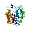
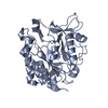
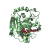
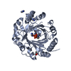
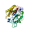
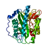
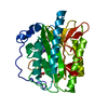
| Title | PHOSPHATIDYLINOSITOL-SPECIFIC PHOSPHOLIPASE C IN COMPLEX WITH MYO-INOSITOL | ||||||
 Components Components | PHOSPHATIDYLINOSITOL-SPECIFIC PHOSPHOLIPASE C | ||||||
 Keywords Keywords | HYDROLASE (PHOSPHORIC DIESTER) / HYDROLASE / PHOSPHATIDYLINOSITOL SPECIFIC PHOSPHOLIPASE C / MYO-INOSITOL / INHIBITOR COMPLEX | ||||||
| Function / homology |  Function and homology information Function and homology informationphosphatidylinositol diacylglycerol-lyase / phosphatidylinositol diacylglycerol-lyase activity / phosphoric diester hydrolase activity / lipid catabolic process / extracellular region Similarity search - Function | ||||||
| Biological species |  | ||||||
| Method |  X-RAY DIFFRACTION / MIR / Resolution: 2.6 Å X-RAY DIFFRACTION / MIR / Resolution: 2.6 Å | ||||||
 Authors Authors | Heinz, D.W. / Ryan, M. / Bullock, T.L. / Griffith, O.H. | ||||||
 Citation Citation | Journal: EMBO J. / Year: 1995 Title: Crystal structure of the phosphatidylinositol-specific phospholipase C from Bacillus cereus in complex with myo-inositol. Authors: Heinz, D.W. / Ryan, M. / Bullock, T.L. / Griffith, O.H. #1: Journal: Biophys.J. / Year: 1993Title: Crystallization of Phosphatidylinositol-Specific Phospholipase C from Bacillus Cereus Authors: Bullock, T.L. / Ryan, M. / Kim, S.L. / Remington, S.J. / Griffith, O.H. #2: Journal: J.Bacteriol. / Year: 1989Title: Phosphatidylinositol-Specific Phospholipase C of Bacillus Cereus: Cloning, Sequencing, and Relationship to Other Phospholipases Authors: Kuppe, A. / Evans, L.M. / Mcmillen, D.A. / Griffith, O.H. | ||||||
| History |
|
- Structure visualization
| Structure viewer | Molecule: MolmilJmol/JSmol |
|---|
- Downloads & links
-Download
| PDBx/mmCIF format | 1ptg.cif.gz | 71.9 KB | Display | PDBx/mmCIF format |
|---|---|---|---|---|
| PDB format | pdb1ptg.ent.gz | 54.3 KB | Display | PDB format |
| PDBx/mmJSON format | 1ptg.json.gz | Tree view | PDBx/mmJSON format | |
| Others |  Other downloads Other downloads |
-Validation report
| Arichive directory | https://data.pdbj.org/pub/pdb/validation_reports/pt/1ptgftp://data.pdbj.org/pub/pdb/validation_reports/pt/1ptg | HTTPS FTP |
|---|
-Related structure data


-Links
 PDBj
PDBj- Assembly
| Deposited unit | 
| ||||||||
|---|---|---|---|---|---|---|---|---|---|
| 1 |
| ||||||||
| Unit cell |
|
-Components
| #1: Protein | Mass: 34568.664 Da / Num. of mol.: 1 Source method: isolated from a genetically manipulated source Source: (gene. exp.) |
|---|---|
| #2: Chemical | ChemComp-INS /   Mass: 180.156 Da / Num. of mol.: 1 / Source method: obtained synthetically / Formula: C6H12O6 Mass: 180.156 Da / Num. of mol.: 1 / Source method: obtained synthetically / Formula: C6H12O6 |
| #3: Water | ChemComp-HOH /  Mass: 18.015 Da / Num. of mol.: 53 / Source method: isolated from a natural source / Formula: H2O Mass: 18.015 Da / Num. of mol.: 53 / Source method: isolated from a natural source / Formula: H2O |
-Experimental details
-Experiment
| Experiment | Method: X-RAY DIFFRACTION |
|---|
- Sample preparation
| Crystal | Density Matthews: 2.4 Å3/Da / Density % sol: 45 % |
|---|
-Data collection
| Diffraction source | Wavelength: 1.5418 |
|---|---|
| Detector | Type: SIEMENS / Detector: AREA DETECTOR / Date: 1994 |
| Radiation | Monochromatic (M) / Laue (L): M / Scattering type: x-ray |
| Radiation wavelength | Wavelength: 1.5418 Å / Relative weight: 1 |
| Reflection | Resolution: 2.5→20 Å / Num. obs: 10300 / % possible obs: 78 % / Observed criterion σ(I): 1 / Redundancy: 3 % / Rmerge(I) obs: 0.045 |
- Processing
| Software |
| ||||||||||||||||||||||||||||||||||||||||||||||||||||||||||||
|---|---|---|---|---|---|---|---|---|---|---|---|---|---|---|---|---|---|---|---|---|---|---|---|---|---|---|---|---|---|---|---|---|---|---|---|---|---|---|---|---|---|---|---|---|---|---|---|---|---|---|---|---|---|---|---|---|---|---|---|---|---|
| Refinement | Method to determine structure: MIR / Resolution: 2.6→8 Å / σ(F): 1
| ||||||||||||||||||||||||||||||||||||||||||||||||||||||||||||
| Displacement parameters | Biso mean: 19.5 Å2 | ||||||||||||||||||||||||||||||||||||||||||||||||||||||||||||
| Refine analyze | Luzzati coordinate error obs: 0.32 Å | ||||||||||||||||||||||||||||||||||||||||||||||||||||||||||||
| Refinement step | Cycle: LAST / Resolution: 2.6→8 Å
| ||||||||||||||||||||||||||||||||||||||||||||||||||||||||||||
| Refine LS restraints |
|
