 Movie
Movie Controller

[English] 日本語
 Yorodumi
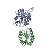
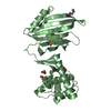
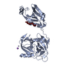
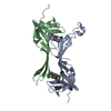
- PDB-1poe: STRUCTURES OF FREE AND INHIBITED HUMAN SECRETORY PHOSPHOLIPASE A2... -
+ Open data

- Basic information
| Entry | Database: PDB / ID: 1poe | ||||||
|---|---|---|---|---|---|---|---|
| Title | STRUCTURES OF FREE AND INHIBITED HUMAN SECRETORY PHOSPHOLIPASE A2 FROM INFLAMMATORY EXUDATE | ||||||
 Components Components | PHOSPHOLIPASE A2 | ||||||
 Keywords Keywords | HYDROLASE | ||||||
| Function / homology |  Function and homology information Function and homology informationregulation of neutrophil activation / phosphatidylethanolamine metabolic process / phosphatidic acid metabolic process / Acyl chain remodelling of PG / Acyl chain remodelling of PC / Acyl chain remodelling of PI / Acyl chain remodelling of PS / Acyl chain remodelling of PE / intestinal stem cell homeostasis / Synthesis of PA ...regulation of neutrophil activation / phosphatidylethanolamine metabolic process / phosphatidic acid metabolic process / Acyl chain remodelling of PG / Acyl chain remodelling of PC / Acyl chain remodelling of PI / Acyl chain remodelling of PS / Acyl chain remodelling of PE / intestinal stem cell homeostasis / Synthesis of PA / phosphatidylglycerol metabolic process / phosphatidylcholine metabolic process / A2-type glycerophospholipase activity / phospholipase A2 / low-density lipoprotein particle remodeling / positive regulation of macrophage derived foam cell differentiation / Antimicrobial peptides / arachidonate secretion / phospholipid metabolic process / negative regulation of T cell proliferation / lipid catabolic process / secretory granule / angiotensin-activated signaling pathway / phospholipid binding / positive regulation of inflammatory response / killing of cells of another organism / positive regulation of ERK1 and ERK2 cascade / mitochondrial outer membrane / defense response to Gram-positive bacterium / inflammatory response / calcium ion binding / endoplasmic reticulum membrane / perinuclear region of cytoplasm / endoplasmic reticulum / : / extracellular exosome / extracellular region / plasma membrane Similarity search - Function | ||||||
| Biological species |  Homo sapiens (human) Homo sapiens (human) | ||||||
| Method |  X-RAY DIFFRACTION / Resolution: 2.1 Å X-RAY DIFFRACTION / Resolution: 2.1 Å | ||||||
 Authors Authors | Scott, D.L. / White, S.P. / Sigler, P.B. | ||||||
 Citation Citation | Journal: Science / Year: 1991 Title: Structures of free and inhibited human secretory phospholipase A2 from inflammatory exudate. Authors: Scott, D.L. / White, S.P. / Browning, J.L. / Rosa, J.J. / Gelb, M.H. / Sigler, P.B. #1: Journal: Science / Year: 1990Title: Interfacial Catalysis: The Mechanism of Phospholipase A2 Authors: Scott, D.L. / White, S.P. / Otwinowski, Z. / Yuan, W. / Gelb, M.H. / Sigler, P.B. | ||||||
| History |
|
- Structure visualization
| Structure viewer | Molecule: MolmilJmol/JSmol |
|---|
- Downloads & links
-Download
| PDBx/mmCIF format | 1poe.cif.gz | 71.4 KB | Display | PDBx/mmCIF format |
|---|---|---|---|---|
| PDB format | pdb1poe.ent.gz | 53.1 KB | Display | PDB format |
| PDBx/mmJSON format | 1poe.json.gz | Tree view | PDBx/mmJSON format | |
| Others |  Other downloads Other downloads |
-Validation report
| Arichive directory | https://data.pdbj.org/pub/pdb/validation_reports/po/1poeftp://data.pdbj.org/pub/pdb/validation_reports/po/1poe | HTTPS FTP |
|---|
-Related structure data


-Links
 PDBj
PDBj


- Assembly
| Deposited unit | 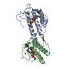
| ||||||||
|---|---|---|---|---|---|---|---|---|---|
| 1 |
| ||||||||
| Unit cell |
|
-Components
| #1: Protein | Mass: 13945.012 Da / Num. of mol.: 2 Source method: isolated from a genetically manipulated source Source: (gene. exp.) Homo sapiens (human) / References: UniProt: P14555, phospholipase A2#2: Chemical | ChemComp-CA /   Mass: 40.078 Da / Num. of mol.: 4 / Source method: obtained synthetically / Formula: Ca Mass: 40.078 Da / Num. of mol.: 4 / Source method: obtained synthetically / Formula: Ca#3: Chemical | 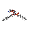  Mass: 489.521 Da / Num. of mol.: 2 / Source method: obtained synthetically / Formula: C20H45NO8P2 Mass: 489.521 Da / Num. of mol.: 2 / Source method: obtained synthetically / Formula: C20H45NO8P2#4: Water | ChemComp-HOH / |  Mass: 18.015 Da / Num. of mol.: 314 / Source method: isolated from a natural source / Formula: H2O Mass: 18.015 Da / Num. of mol.: 314 / Source method: isolated from a natural source / Formula: H2OHas protein modification | Y | |
|---|
-Experimental details
-Experiment
| Experiment | Method: X-RAY DIFFRACTION |
|---|
- Sample preparation
| Crystal | Density Matthews: 3.01 Å3/Da / Density % sol: 59.11 % | ||||||||||||||||||||||||||||||||||||||||||
|---|---|---|---|---|---|---|---|---|---|---|---|---|---|---|---|---|---|---|---|---|---|---|---|---|---|---|---|---|---|---|---|---|---|---|---|---|---|---|---|---|---|---|---|
| Crystal grow | *PLUS pH: 7.4 / Method: vapor diffusion | ||||||||||||||||||||||||||||||||||||||||||
| Components of the solutions | *PLUS
|
-Data collection
| Radiation | Scattering type: x-ray |
|---|---|
| Radiation wavelength | Relative weight: 1 |
- Processing
| Software |
| ||||||||||||||||||||||||||||||||||||||||||||||||||||||||||||
|---|---|---|---|---|---|---|---|---|---|---|---|---|---|---|---|---|---|---|---|---|---|---|---|---|---|---|---|---|---|---|---|---|---|---|---|---|---|---|---|---|---|---|---|---|---|---|---|---|---|---|---|---|---|---|---|---|---|---|---|---|---|
| Refinement | Rfactor Rwork: 0.196 / Rfactor obs: 0.196 / Highest resolution: 2.1 Å | ||||||||||||||||||||||||||||||||||||||||||||||||||||||||||||
| Refinement step | Cycle: LAST / Highest resolution: 2.1 Å
| ||||||||||||||||||||||||||||||||||||||||||||||||||||||||||||
| Refine LS restraints |
| ||||||||||||||||||||||||||||||||||||||||||||||||||||||||||||
| Software | *PLUS Name: X-PLOR / Classification: refinement | ||||||||||||||||||||||||||||||||||||||||||||||||||||||||||||
| Refinement | *PLUS Highest resolution: 2.1 Å / Rfactor obs: 0.196 | ||||||||||||||||||||||||||||||||||||||||||||||||||||||||||||
| Solvent computation | *PLUS | ||||||||||||||||||||||||||||||||||||||||||||||||||||||||||||
| Displacement parameters | *PLUS | ||||||||||||||||||||||||||||||||||||||||||||||||||||||||||||
| Refine LS restraints | *PLUS Type: x_angle_d / Dev ideal: 2.2 |
