 Movie
Movie Controller

[English] 日本語
 Yorodumi
- PDB-1pnq: Crystal structure of R. rubrum transhydrogenase domain III bound ... -
+ Open data

- Basic information
| Entry | Database: PDB / ID: 1pnq | ||||||
|---|---|---|---|---|---|---|---|
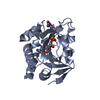
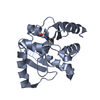
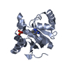
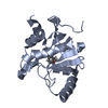
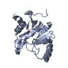
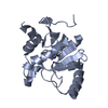
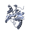
| Title | Crystal structure of R. rubrum transhydrogenase domain III bound to NADPH | ||||||
 Components Components | NAD(P) transhydrogenase subunit beta | ||||||
 Keywords Keywords | OXIDOREDUCTASE / Nucleotide binding fold / NADPH | ||||||
| Function / homology |  Function and homology information Function and homology informationproton-translocating NAD(P)+ transhydrogenase activity / proton-translocating NAD(P)+ transhydrogenase / membrane => GO:0016020 / NADP binding / plasma membrane Similarity search - Function | ||||||
| Biological species |  Rhodospirillum rubrum (bacteria) Rhodospirillum rubrum (bacteria) | ||||||
| Method |  X-RAY DIFFRACTION / SYNCHROTRON / MOLECULAR REPLACEMENT / Resolution: 2.4 Å X-RAY DIFFRACTION / SYNCHROTRON / MOLECULAR REPLACEMENT / Resolution: 2.4 Å | ||||||
 Authors Authors | Sundaresan, V. / Yamaguchi, M. / Chartron, J. / Stout, C.D. | ||||||
 Citation Citation | Journal: Biochemistry / Year: 2003 Title: Conformational Change in the NADP(H) Binding Domain of Transhydrogenase Defines Four States Authors: Sundaresan, V. / Yamaguchi, M. / Chartron, J. / Stout, C.D. | ||||||
| History |
|
- Structure visualization
| Structure viewer | Molecule: MolmilJmol/JSmol |
|---|
- Downloads & links
-Download
| PDBx/mmCIF format | 1pnq.cif.gz | 85.3 KB | Display | PDBx/mmCIF format |
|---|---|---|---|---|
| PDB format | pdb1pnq.ent.gz | 65.7 KB | Display | PDB format |
| PDBx/mmJSON format | 1pnq.json.gz | Tree view | PDBx/mmJSON format | |
| Others |  Other downloads Other downloads |
-Validation report
| Arichive directory | https://data.pdbj.org/pub/pdb/validation_reports/pn/1pnqftp://data.pdbj.org/pub/pdb/validation_reports/pn/1pnq | HTTPS FTP |
|---|
-Related structure data


-Links
 PDBj
PDBj


- Assembly
| Deposited unit | 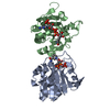
| |||||||||
|---|---|---|---|---|---|---|---|---|---|---|
| 1 | 
| |||||||||
| 2 | 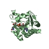
| |||||||||
| Unit cell |
| |||||||||
| Components on special symmetry positions |
|
-Components
| #1: Protein | Mass: 19442.213 Da / Num. of mol.: 2 Source method: isolated from a genetically manipulated source Source: (gene. exp.) Rhodospirillum rubrum (bacteria) / Production host: References: UniProt: Q59765, UniProt: Q2RSB4*PLUS, EC: 1.6.1.2 #2: Chemical |   Mass: 745.421 Da / Num. of mol.: 2 / Source method: obtained synthetically / Formula: C21H30N7O17P3 Mass: 745.421 Da / Num. of mol.: 2 / Source method: obtained synthetically / Formula: C21H30N7O17P3#3: Water | ChemComp-HOH / |  Mass: 18.015 Da / Num. of mol.: 165 / Source method: isolated from a natural source / Formula: H2O Mass: 18.015 Da / Num. of mol.: 165 / Source method: isolated from a natural source / Formula: H2O |
|---|
-Experimental details
-Experiment
| Experiment | Method: X-RAY DIFFRACTION / Number of used crystals: 1 |
|---|
- Sample preparation
| Crystal grow | Temperature: 281 K / Method: vapor diffusion, hanging drop / pH: 5.5 Details: ammonium sulfate, citrate, pH 5.5, VAPOR DIFFUSION, HANGING DROP, temperature 281K | ||||||||||||||||||||||||||||||||||||||||||||||||
|---|---|---|---|---|---|---|---|---|---|---|---|---|---|---|---|---|---|---|---|---|---|---|---|---|---|---|---|---|---|---|---|---|---|---|---|---|---|---|---|---|---|---|---|---|---|---|---|---|---|
| Crystal grow | *PLUS Temperature: 8 ℃ / Method: vapor diffusion | ||||||||||||||||||||||||||||||||||||||||||||||||
| Components of the solutions | *PLUS
|
-Data collection
| Diffraction | Mean temperature: 100 K |
|---|---|
| Diffraction source | Source: SYNCHROTRON / Site: SSRL  / Beamline: BL9-1 / Wavelength: 0.979 Å / Beamline: BL9-1 / Wavelength: 0.979 Å |
| Detector | Type: ADSC QUANTUM 315 / Detector: CCD / Date: Jan 22, 2002 |
| Radiation | Protocol: SINGLE WAVELENGTH / Monochromatic (M) / Laue (L): M / Scattering type: x-ray |
| Radiation wavelength | Wavelength: 0.979 Å / Relative weight: 1 |
| Reflection | Resolution: 2.4→19.65 Å / Num. obs: 34800 / % possible obs: 99 % / Observed criterion σ(I): 1 / Redundancy: 9.1 % / Biso Wilson estimate: 50.4 Å2 / Rmerge(I) obs: 0.094 / Rsym value: 0.085 / Net I/σ(I): 17.3 |
| Reflection shell | Resolution: 2.4→2.46 Å / Redundancy: 5.1 % / Num. unique all: 2367 / % possible all: 99 |
| Reflection | *PLUS Lowest resolution: 19.7 Å / Num. measured all: 317639 / Rmerge(I) obs: 0.085 |
| Reflection shell | *PLUS Rmerge(I) obs: 0.635 / Mean I/σ(I) obs: 1.4 |
- Processing
| Software |
| ||||||||||||||||||||||||||||||||||||
|---|---|---|---|---|---|---|---|---|---|---|---|---|---|---|---|---|---|---|---|---|---|---|---|---|---|---|---|---|---|---|---|---|---|---|---|---|---|
| Refinement | Method to determine structure: MOLECULAR REPLACEMENT / Resolution: 2.4→19.63 Å / Rfactor Rfree error: 0.006 / Data cutoff high absF: 2344125.44 / Data cutoff high rms absF: 2344125.44 / Data cutoff low absF: 0 / Isotropic thermal model: RESTRAINED / Cross valid method: THROUGHOUT / σ(F): 0
| ||||||||||||||||||||||||||||||||||||
| Solvent computation | Solvent model: FLAT MODEL / Bsol: 41.751 Å2 / ksol: 0.342248 e/Å3 | ||||||||||||||||||||||||||||||||||||
| Displacement parameters | Biso mean: 58.9 Å2
| ||||||||||||||||||||||||||||||||||||
| Refine analyze |
| ||||||||||||||||||||||||||||||||||||
| Refinement step | Cycle: LAST / Resolution: 2.4→19.63 Å
| ||||||||||||||||||||||||||||||||||||
| Refine LS restraints |
| ||||||||||||||||||||||||||||||||||||
| LS refinement shell | Resolution: 2.4→2.55 Å / Rfactor Rfree error: 0.02 / Total num. of bins used: 6
| ||||||||||||||||||||||||||||||||||||
| Xplor file |
| ||||||||||||||||||||||||||||||||||||
| Refinement | *PLUS Highest resolution: 2.4 Å / Lowest resolution: 19.7 Å / % reflection Rfree: 5 % / Rfactor Rfree: 0.231 / Rfactor Rwork: 0.217 | ||||||||||||||||||||||||||||||||||||
| Solvent computation | *PLUS | ||||||||||||||||||||||||||||||||||||
| Displacement parameters | *PLUS | ||||||||||||||||||||||||||||||||||||
| Refine LS restraints | *PLUS
|
