 Movie
Movie Controller

[English] 日本語
 Yorodumi
- PDB-1pa3: Crystal Structure of Glutathione-S-transferase from Plasmodium fa... -
+ Open data

- Basic information
| Entry | Database: PDB / ID: 1pa3 | ||||||
|---|---|---|---|---|---|---|---|
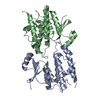
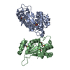
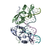
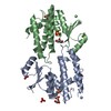
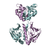
| Title | Crystal Structure of Glutathione-S-transferase from Plasmodium falciparum | ||||||
 Components Components | Glutathione s-transferase, putative | ||||||
 Keywords Keywords | TRANSFERASE | ||||||
| Function / homology |  Function and homology information Function and homology informationGlutathione conjugation / Heme degradation / Paracetamol ADME / Synthesis of Prostaglandins (PG) and Thromboxanes (TX) / Azathioprine ADME / Detoxification of Reactive Oxygen Species / Neutrophil degranulation / glutathione transferase / glutathione transferase activity / glutathione metabolic process Similarity search - Function | ||||||
| Biological species |  | ||||||
| Method |  X-RAY DIFFRACTION / MIR / Resolution: 2.7 Å X-RAY DIFFRACTION / MIR / Resolution: 2.7 Å | ||||||
 Authors Authors | Perbandt, M. / Betzel, C. / Liebau, E. | ||||||
 Citation Citation | Journal: J.Biol.Chem. / Year: 2004 Title: Native and inhibited structure of a Mu class-related glutathione S-transferase from Plasmodium falciparum Authors: Perbandt, M. / Burmeister, C. / Walter, R.D. / Betzel, C. / Liebau, E. | ||||||
| History |
|
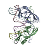
- Structure visualization
| Structure viewer | Molecule: MolmilJmol/JSmol |
|---|
- Downloads & links
-Download
| PDBx/mmCIF format | 1pa3.cif.gz | 90.1 KB | Display | PDBx/mmCIF format |
|---|---|---|---|---|
| PDB format | pdb1pa3.ent.gz | 70.3 KB | Display | PDB format |
| PDBx/mmJSON format | 1pa3.json.gz | Tree view | PDBx/mmJSON format | |
| Others |  Other downloads Other downloads |
-Validation report
| Summary document | 1pa3_validation.pdf.gz | 434.1 KB | Display | wwPDB validaton report |
|---|---|---|---|---|
| Full document | 1pa3_full_validation.pdf.gz | 445 KB | Display | |
| Data in XML | 1pa3_validation.xml.gz | 16.7 KB | Display | |
| Data in CIF | 1pa3_validation.cif.gz | 22.3 KB | Display | |
| Arichive directory | https://data.pdbj.org/pub/pdb/validation_reports/pa/1pa3ftp://data.pdbj.org/pub/pdb/validation_reports/pa/1pa3 | HTTPS FTP |
-Related structure data


-Links
 PDBj
PDBj

- Assembly
| Deposited unit | 
| ||||||||
|---|---|---|---|---|---|---|---|---|---|
| 1 |
| ||||||||
| 2 | 
| ||||||||
| Unit cell |
|
-Components
| #1: Protein | Mass: 24817.039 Da / Num. of mol.: 2 Source method: isolated from a genetically manipulated source Source: (gene. exp.) Production host:  #2: Water | ChemComp-HOH / |  Mass: 18.015 Da / Num. of mol.: 32 / Source method: isolated from a natural source / Formula: H2O Mass: 18.015 Da / Num. of mol.: 32 / Source method: isolated from a natural source / Formula: H2O |
|---|
-Experimental details
-Experiment
| Experiment | Method: X-RAY DIFFRACTION |
|---|
- Sample preparation
| Crystal | Density Matthews: 2.08 Å3/Da / Density % sol: 40.85 % | ||||||||||||||||||||||||||||||
|---|---|---|---|---|---|---|---|---|---|---|---|---|---|---|---|---|---|---|---|---|---|---|---|---|---|---|---|---|---|---|---|
| Crystal grow | Temperature: 298 K / Method: vapor diffusion, hanging drop / pH: 8.5 Details: Ammonium sulfate, Tris, pH 8.5, VAPOR DIFFUSION, HANGING DROP, temperature 298K | ||||||||||||||||||||||||||||||
| Crystal grow | *PLUS pH: 6 / Method: vapor diffusion, hanging drop / Details: Burmeister, C., (2003) Acta Cryst., D59, 1469. | ||||||||||||||||||||||||||||||
| Components of the solutions | *PLUS
|
-Data collection
| Diffraction source | Source: ROTATING ANODE / Type: RIGAKU RU200 / Wavelength: 1.5418 |
|---|---|
| Detector | Type: MARRESEARCH / Detector: IMAGE PLATE / Date: Mar 20, 2003 |
| Radiation | Protocol: SINGLE WAVELENGTH / Monochromatic (M) / Laue (L): M / Scattering type: x-ray |
| Radiation wavelength | Wavelength: 1.5418 Å / Relative weight: 1 |
| Reflection | Resolution: 2.6→50 Å / Num. obs: 13330 / Observed criterion σ(I): 0 |
| Reflection shell | Highest resolution: 2.6 Å / % possible all: 97.92 |
| Reflection | *PLUS Num. obs: 13027 / % possible obs: 97.6 % / Redundancy: 6 % / Rmerge(I) obs: 0.13 |
| Reflection shell | *PLUS Lowest resolution: 2.69 Å / % possible obs: 94.7 % / Redundancy: 5.6 % / Rmerge(I) obs: 0.519 / Mean I/σ(I) obs: 2.3 |
- Processing
| Software | Name: REFMAC / Version: 5 / Classification: refinement | ||||||||||||||||||||
|---|---|---|---|---|---|---|---|---|---|---|---|---|---|---|---|---|---|---|---|---|---|
| Refinement | Method to determine structure: MIR / Resolution: 2.7→15 Å / Cross valid method: THROUGHOUT / σ(F): 0 / Stereochemistry target values: Engh & Huber
| ||||||||||||||||||||
| Refinement step | Cycle: LAST / Resolution: 2.7→15 Å
| ||||||||||||||||||||
| Software | *PLUS Name: REFMAC / Version: 5 / Classification: refinement | ||||||||||||||||||||
| Refinement | *PLUS Highest resolution: 2.6 Å / Num. reflection Rfree: 658 / % reflection Rfree: 5 % / Rfactor Rfree: 0.258 / Rfactor Rwork: 0.205 | ||||||||||||||||||||
| Solvent computation | *PLUS | ||||||||||||||||||||
| Displacement parameters | *PLUS | ||||||||||||||||||||
| Refine LS restraints | *PLUS
| ||||||||||||||||||||
| LS refinement shell | *PLUS Rfactor Rfree: 0.351 / Rfactor Rwork: 0.322 |
