 Movie
Movie Controller

[English] 日本語
 Yorodumi
- PDB-1p9b: Structure of fully ligated Adenylosuccinate synthetase from Plasm... -
+ Open data

- Basic information
| Entry | Database: PDB / ID: 1p9b | ||||||
|---|---|---|---|---|---|---|---|
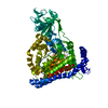
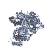
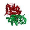
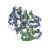
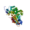
| Title | Structure of fully ligated Adenylosuccinate synthetase from Plasmodium falciparum | ||||||
 Components Components | Adenylosuccinate Synthetase | ||||||
 Keywords Keywords | LIGASE | ||||||
| Function / homology |  Function and homology information Function and homology informationadenylosuccinate synthase / adenylosuccinate synthase activity / IMP metabolic process / 'de novo' AMP biosynthetic process / GTP binding / magnesium ion binding / cytoplasm Similarity search - Function | ||||||
| Biological species |  | ||||||
| Method |  X-RAY DIFFRACTION / MOLECULAR REPLACEMENT / Resolution: 2 Å X-RAY DIFFRACTION / MOLECULAR REPLACEMENT / Resolution: 2 Å | ||||||
 Authors Authors | Eaazhisai, K. / Jayalakshmi, R. / Gayathri, P. / Anand, R.P. / Sumathy, K. / Balaram, H. / Murthy, M.R. | ||||||
 Citation Citation | Journal: J.Mol.Biol. / Year: 2004 Title: Crystal Structure of Fully Ligated Adenylosuccinate Synthetase from Plasmodium falciparum. Authors: Eaazhisai, K. / Jayalakshmi, R. / Gayathri, P. / Anand, R.P. / Sumathy, K. / Balaram, H. / Murthy, M.R. | ||||||
| History |
|
- Structure visualization
| Structure viewer | Molecule: MolmilJmol/JSmol |
|---|
- Downloads & links
-Download
| PDBx/mmCIF format | 1p9b.cif.gz | 107.3 KB | Display | PDBx/mmCIF format |
|---|---|---|---|---|
| PDB format | pdb1p9b.ent.gz | 79.7 KB | Display | PDB format |
| PDBx/mmJSON format | 1p9b.json.gz | Tree view | PDBx/mmJSON format | |
| Others |  Other downloads Other downloads |
-Validation report
| Arichive directory | https://data.pdbj.org/pub/pdb/validation_reports/p9/1p9bftp://data.pdbj.org/pub/pdb/validation_reports/p9/1p9b | HTTPS FTP |
|---|
-Related structure data
| Related structure data | 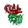 1cibS S: Starting model for refinement |
|---|---|
| Similar structure data |


-Links
 PDBj
PDBj- Assembly
| Deposited unit | 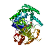
| ||||||||
|---|---|---|---|---|---|---|---|---|---|
| 1 | 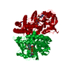
| ||||||||
| Unit cell |
| ||||||||
| Details | The second part of the biological assembly is generated by the two fold axis: -X,Y,1/2-Z |
-Components
-Protein , 1 types, 1 molecules A
| #1: Protein | Mass: 50088.738 Da / Num. of mol.: 1 Source method: isolated from a genetically manipulated source Source: (gene. exp.) Plasmid: pET23d / Species (production host): Escherichia coli / Production host:  |
|---|
-Non-polymers , 6 types, 283 molecules 


| #2: Chemical | ChemComp-MG /  Mass: 24.305 Da / Num. of mol.: 1 / Source method: obtained synthetically / Formula: Mg Mass: 24.305 Da / Num. of mol.: 1 / Source method: obtained synthetically / Formula: Mg |
|---|---|
| #3: Chemical | ChemComp-NO3 /  Mass: 62.005 Da / Num. of mol.: 1 / Source method: obtained synthetically / Formula: NO3 Mass: 62.005 Da / Num. of mol.: 1 / Source method: obtained synthetically / Formula: NO3 |
| #4: Chemical | ChemComp-IMO /  Mass: 428.186 Da / Num. of mol.: 1 / Source method: obtained synthetically / Formula: C10H14N4O11P2 Mass: 428.186 Da / Num. of mol.: 1 / Source method: obtained synthetically / Formula: C10H14N4O11P2 |
| #5: Chemical | ChemComp-HDA /  Mass: 119.076 Da / Num. of mol.: 1 / Source method: obtained synthetically / Formula: C3H5NO4 / Comment: anticancer, medication*YM Mass: 119.076 Da / Num. of mol.: 1 / Source method: obtained synthetically / Formula: C3H5NO4 / Comment: anticancer, medication*YM |
| #6: Chemical | ChemComp-GDP /  Type: RNA linking / Mass: 443.201 Da / Num. of mol.: 1 / Source method: obtained synthetically / Formula: C10H15N5O11P2 / Comment: GDP, energy-carrying molecule*YM Type: RNA linking / Mass: 443.201 Da / Num. of mol.: 1 / Source method: obtained synthetically / Formula: C10H15N5O11P2 / Comment: GDP, energy-carrying molecule*YM |
| #7: Water | ChemComp-HOH / Mass: 18.015 Da / Num. of mol.: 278 / Source method: isolated from a natural source / Formula: H2O |
-Experimental details
-Experiment
| Experiment | Method: X-RAY DIFFRACTION / Number of used crystals: 1 |
|---|
- Sample preparation
| Crystal | Density Matthews: 2.08 Å3/Da / Density % sol: 40.49 % | ||||||||||||||||||||||||||||||||||||
|---|---|---|---|---|---|---|---|---|---|---|---|---|---|---|---|---|---|---|---|---|---|---|---|---|---|---|---|---|---|---|---|---|---|---|---|---|---|
| Crystal grow | Temperature: 293 K / Method: liquid diffusion / pH: 6.5 Details: PEG4000, Sodium cacodylate, pH 6.5, LIQUID DIFFUSION, temperature 293K | ||||||||||||||||||||||||||||||||||||
| Crystal grow | *PLUS Method: vapor diffusion, hanging drop | ||||||||||||||||||||||||||||||||||||
| Components of the solutions | *PLUS
|
-Data collection
| Diffraction | Mean temperature: 110 K |
|---|---|
| Diffraction source | Source: ROTATING ANODE / Type: RIGAKU RU200 / Wavelength: 1.5418 Å |
| Detector | Type: MARRESEARCH / Detector: IMAGE PLATE / Date: Mar 10, 2003 / Details: mirrors |
| Radiation | Protocol: SINGLE WAVELENGTH / Monochromatic (M) / Laue (L): M / Scattering type: x-ray |
| Radiation wavelength | Wavelength: 1.5418 Å / Relative weight: 1 |
| Reflection | Resolution: 2→32.91 Å / Num. all: 164288 / Num. obs: 28913 / Redundancy: 5.7 % / Biso Wilson estimate: 17.2 Å2 / Rmerge(I) obs: 0.075 / Rsym value: 0.068 / Net I/σ(I): 7.2 |
| Reflection shell | Resolution: 2→2.1 Å / Redundancy: 5.5 % / Rmerge(I) obs: 0.397 / Mean I/σ(I) obs: 2.2 / Num. unique all: 2833 / Rsym value: 0.36 |
| Reflection | *PLUS Lowest resolution: 50 Å / Num. obs: 29594 / % possible obs: 97.8 % / Num. measured all: 358593 / Rmerge(I) obs: 0.068 |
| Reflection shell | *PLUS Highest resolution: 2 Å / Lowest resolution: 2.07 Å / % possible obs: 97.1 % / Rmerge(I) obs: 0.363 |
- Processing
| Software |
| |||||||||||||||||||||||||
|---|---|---|---|---|---|---|---|---|---|---|---|---|---|---|---|---|---|---|---|---|---|---|---|---|---|---|
| Refinement | Method to determine structure: MOLECULAR REPLACEMENT Starting model: PDB ENTRY 1CIB Resolution: 2→32.91 Å / Isotropic thermal model: Anisotropic / Cross valid method: THROUGHOUT / Stereochemistry target values: Engh & Huber
| |||||||||||||||||||||||||
| Displacement parameters | Biso mean: 25.2 Å2
| |||||||||||||||||||||||||
| Refine analyze |
| |||||||||||||||||||||||||
| Refinement step | Cycle: LAST / Resolution: 2→32.91 Å
| |||||||||||||||||||||||||
| Refine LS restraints |
| |||||||||||||||||||||||||
| LS refinement shell | Resolution: 2→2.13 Å / Rfactor Rfree error: 0.025
| |||||||||||||||||||||||||
| Refinement | *PLUS Highest resolution: 2 Å / Lowest resolution: 50 Å | |||||||||||||||||||||||||
| Solvent computation | *PLUS | |||||||||||||||||||||||||
| Displacement parameters | *PLUS | |||||||||||||||||||||||||
| Refine LS restraints | *PLUS
|
