 ムービー
ムービー コントローラー


+ データを開く

- 基本情報
基本情報
| 登録情報 | データベース: PDB / ID: 1p5c | ||||||
|---|---|---|---|---|---|---|---|
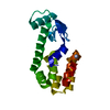
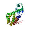
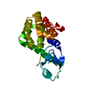
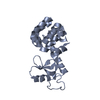
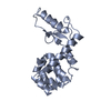
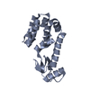
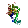
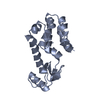
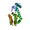
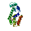
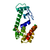
| タイトル | Circular permutation of Helix A in T4 lysozyme | ||||||
 要素 要素 | Lysozyme | ||||||
 キーワード キーワード | HYDROLASE / Circular permutation / protein design / context dependent folding | ||||||
| 機能・相同性 |  機能・相同性情報 機能・相同性情報viral release from host cell by cytolysis / peptidoglycan catabolic process / cell wall macromolecule catabolic process / lysozyme / lysozyme activity / host cell cytoplasm / defense response to bacterium 類似検索 - 分子機能 | ||||||
| 生物種 |  Enterobacteria phage T4 (ファージ) Enterobacteria phage T4 (ファージ) | ||||||
| 手法 |  X線回折 / シンクロトロン / 分子置換 / 解像度: 2.5 Å X線回折 / シンクロトロン / 分子置換 / 解像度: 2.5 Å | ||||||
 データ登録者 データ登録者 | Sagermann, M. / Gay, L. / Baase, W.A. / Matthews, B.W. | ||||||
 引用 引用 | ジャーナル: Biochemistry / 年: 2004 タイトル: Relocation or duplication of the helix A sequence of T4 lysozyme causes only modest changes in structure but can increase or decrease the rate of folding. 著者: Sagermann, M. / Baase, W.A. / Mooers, B.H. / Gay, L. / Matthews, B.W. #1: ジャーナル: J.Mol.Biol. / 年: 2002タイトル: Crystal structures of a T4-lysozyme duplication-extension mutant demonstrates that the highly conserved beta-sheet region has low intrinsic folding propensity 著者: Sagermann, M. / Matthews, B.W. #2: ジャーナル: J.Mol.Biol. / 年: 1987タイトル: Structure of bacteriophage T4 lysozyme refined at 1.7 A resolution 著者: Weaver, L.H. / Matthews, B.W. #3: ジャーナル: Proc.Natl.Acad.Sci.USA / 年: 1999タイトル: Structural characterization of an engineered tandem repeat contrasts the importance of context and sequence in protein folding 著者: Sagermann, M. / Baase, W.A. / Matthews, B.W. | ||||||
| 履歴 |
| ||||||
| Remark 999 | SEQUENCE Residue Leu 164 was deleted and sequence SGGAMNIFEMLRIDE was appended to the C-terminus |
- 構造の表示
構造の表示
| 構造ビューア | 分子: MolmilJmol/JSmol |
|---|
- ダウンロードとリンク
-ダウンロード
| PDBx/mmCIF形式 | 1p5c.cif.gz | 144.7 KB | 表示 | PDBx/mmCIF形式 |
|---|---|---|---|---|
| PDB形式 | pdb1p5c.ent.gz | 114.7 KB | 表示 | PDB形式 |
| PDBx/mmJSON形式 | 1p5c.json.gz | ツリー表示 | PDBx/mmJSON形式 | |
| その他 |  その他のダウンロード その他のダウンロード |
-検証レポート
| 文書・要旨 | 1p5c_validation.pdf.gz | 453.7 KB | 表示 | wwPDB検証レポート |
|---|---|---|---|---|
| 文書・詳細版 | 1p5c_full_validation.pdf.gz | 469.6 KB | 表示 | |
| XML形式データ | 1p5c_validation.xml.gz | 29.2 KB | 表示 | |
| CIF形式データ | 1p5c_validation.cif.gz | 40.8 KB | 表示 | |
| アーカイブディレクトリ | https://data.pdbj.org/pub/pdb/validation_reports/p5/1p5cftp://data.pdbj.org/pub/pdb/validation_reports/p5/1p5c | HTTPS FTP |
-関連構造データ


-リンク
 PDBj
PDBj


- 集合体
集合体
| 登録構造単位 | 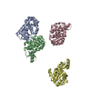
| ||||||||
|---|---|---|---|---|---|---|---|---|---|
| 1 | 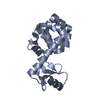
| ||||||||
| 2 | 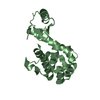
| ||||||||
| 3 | 
| ||||||||
| 4 | 
| ||||||||
| 単位格子 |
| ||||||||
| 詳細 | Protein is a monomer in solution. The asymmetric unit contains 4 molecules. The refinement was performed in the absence of NCS operators. |
-要素
| #1: タンパク質 | 分子量: 18861.607 Da / 分子数: 4 / 変異: C54T, C97A, G12M / 由来タイプ: 組換発現 由来: (組換発現) Enterobacteria phage T4 (ファージ)属: T4-like viruses / 生物種: Enterobacteria phage T4 sensu lato / 遺伝子: GENE E / プラスミド: phs1403 / 発現宿主:  #2: 水 | ChemComp-HOH / |  分子量: 18.015 Da / 分子数: 286 / 由来タイプ: 天然 / 式: H2O 分子量: 18.015 Da / 分子数: 286 / 由来タイプ: 天然 / 式: H2O |
|---|
-実験情報
-実験
| 実験 | 手法: X線回折 / 使用した結晶の数: 1 |
|---|
- 試料調製
試料調製
| 結晶 | マシュー密度: 2.27 Å3/Da / 溶媒含有率: 45.77 % |
|---|---|
| 結晶化 | 温度: 298 K / 手法: 蒸気拡散法, ハンギングドロップ法 / pH: 7.1 詳細: 30% PEG 3400, 50mM Phosphate buffer, 5% isopropanol, pH 7.1, VAPOR DIFFUSION, HANGING DROP, temperature 298K |
-データ収集
| 回折 | 平均測定温度: 170 K |
|---|---|
| 放射光源 | 由来: シンクロトロン / サイト: SSRL  / ビームライン: BL9-1 / 波長: 0.9537 Å / ビームライン: BL9-1 / 波長: 0.9537 Å |
| 検出器 | タイプ: MARRESEARCH / 検出器: IMAGE PLATE / 日付: 2002年4月7日 / 詳細: mirror |
| 放射 | モノクロメーター: single SI crystal / プロトコル: SINGLE WAVELENGTH / 単色(M)・ラウエ(L): M / 散乱光タイプ: x-ray |
| 放射波長 | 波長: 0.9537 Å / 相対比: 1 |
| 反射 | 解像度: 2.5→25 Å / Num. all: 57195 / Num. obs: 22893 / % possible obs: 97.4 % / Observed criterion σ(F): 0 / Observed criterion σ(I): 0 / 冗長度: 2.5 % / Biso Wilson estimate: 35.11 Å2 / Rsym value: 0.076 / Net I/σ(I): 6.7 |
| 反射 シェル | 解像度: 2.5→2.64 Å / 冗長度: 2.4 % / Num. unique all: 3389 / Rsym value: 0.088 / % possible all: 99.3 |
- 解析
解析
| ソフトウェア |
| |||||||||||||||||||||||||
|---|---|---|---|---|---|---|---|---|---|---|---|---|---|---|---|---|---|---|---|---|---|---|---|---|---|---|
| 精密化 | 構造決定の手法: 分子置換 開始モデル: PDB ENTRY 2LZM 解像度: 2.5→25 Å / 交差検証法: THROUGHOUT / σ(F): 0 / 立体化学のターゲット値: Engh & Huber 詳細: Amino terminal Methionine residue not visible in density.
| |||||||||||||||||||||||||
| 原子変位パラメータ |
| |||||||||||||||||||||||||
| 精密化ステップ | サイクル: LAST / 解像度: 2.5→25 Å
| |||||||||||||||||||||||||
| 拘束条件 |
|
