 Movie
Movie Controller

[English] 日本語
 Yorodumi
- PDB-1p5a: Conformational Mapping of the N-terminal Peptide of HIV-1 GP41 in... -
+ Open data

- Basic information
| Entry | Database: PDB / ID: 1p5a | ||||||
|---|---|---|---|---|---|---|---|
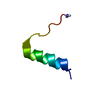
| Title | Conformational Mapping of the N-terminal Peptide of HIV-1 GP41 in lipid detergent and aqueous environments using 13C-enhanced Fourier Transform Infrared Spectroscopy | ||||||
 Components Components | Envelope polyprotein GP160 | ||||||
 Keywords Keywords | VIRAL PROTEIN / human immunodeficiency virus (HIV-1) / viral fusion peptide / gp41 | ||||||
| Function / homology |  Function and homology information Function and homology informationDectin-2 family / symbiont-mediated perturbation of host defense response / positive regulation of plasma membrane raft polarization / positive regulation of receptor clustering / host cell endosome membrane / clathrin-dependent endocytosis of virus by host cell / viral protein processing / fusion of virus membrane with host plasma membrane / fusion of virus membrane with host endosome membrane / viral envelope ...Dectin-2 family / symbiont-mediated perturbation of host defense response / positive regulation of plasma membrane raft polarization / positive regulation of receptor clustering / host cell endosome membrane / clathrin-dependent endocytosis of virus by host cell / viral protein processing / fusion of virus membrane with host plasma membrane / fusion of virus membrane with host endosome membrane / viral envelope / virion attachment to host cell / host cell plasma membrane / virion membrane / structural molecule activity / membrane Similarity search - Function | ||||||
| Method | INFRARED SPECTROSCOPY / molecular dynamics | ||||||
 Authors Authors | Gordon, L.M. / Mobley, P.W. / Lee, W. / Eskandari, S. / Kaznessis, Y.N. / Sherman, M.A. / Waring, A.J. | ||||||
 Citation Citation | Journal: Protein Sci. / Year: 2004 Title: Conformational mapping of the N-terminal peptide of HIV-1 gp41 in lipid detergent and aqueous environments using 13C-enhanced Fourier transform infrared spectroscopy. Authors: Gordon, L.M. / Mobley, P.W. / Lee, W. / Eskandari, S. / Kaznessis, Y.N. / Sherman, M.A. / Waring, A.J. #1: Journal: BIOCHIM.BIOPHYS.ACTA / Year: 2002Title: CONFORMATIONAL MAPPING OF THE N-TERMINAL PEPTIDE OF HIV-1 GP41 IN MEMBRANE ENVIRONMENTS USING 13C-ENHANCED FOURIER TRANSFORM INFRARED SPECTROSCOPY Authors: GORDON, L.M. / MOBLEY, P.W. / PILPA, R. / SHERMAN, M.A. / WARING, A.J. | ||||||
| History |
| ||||||
| Remark 250 | REMARK: THIS STRUCTURE WAS DETERMINED BY MEANS OF 13C-ENHANCED FOURIER TRANSFORM INFRARED ...REMARK: THIS STRUCTURE WAS DETERMINED BY MEANS OF 13C-ENHANCED FOURIER TRANSFORM INFRARED SPECTROSCOPY (FTIR). DATA SET CONSISTS OF 19 CONFORMERS DERIVED FROM MOLECULAR DYNAMICS IN A MODELED SDS MICELLE SURROUNDED BY WATER WITH PEPTIDE BACKBONE CONSTRAINTS FROM FTIR. EXPERIMENTAL DETAILS EXPERIMENT TYPE : INFRARED SPECTROSCOPY TEMPERATURE (KELVIN) : 298 PH : 7.4 IONIC STRENGTH : 0.271 PRESSURE : AMBIENT SAMPLE CONTENTS : THIS STRUCTURE WAS DETERMINED USING 13-C ISOTOPE ENHANCED FTIR SPECTROSCOPY ON A FAMILY OF SELECTIVELY LABELED CHEMICALLY SYNTHESIZED PEPTIDES. 13-C CARBONYL LABELS INCLUDED RESIDUES 519,521,523,524,525,526,527, 528,529,530,531,532,533,534,538,539. EXPERIMENTS CONDUCTED : 13C-ISOTOPE ENHANCED FTIR SPECTROMETER FIELD STRENGTH : NULL SPECTROMETER MODEL : RESEARCH SERIES SPECTROMETER MANUFACTURER : MATTSON FTIR STRUCTURE DETERMINATION. SOFTWARE USED : WINFIRST FTIR CURVE FITTING SOFTWARE METHOD USED : MOLECULAR DYNAMICS CONFORMERS, NUMBER CALCULATED : 20 CONFORMERS, NUMBER SUBMITTED : 19 CONFORMERS, SELECTION CRITERIA : STRUCTURES WITH ACCEPTABLE COVALENT GEOMETRY BEST REPRESENTATIVE CONFORMER IN THIS ENSEMBLE : 1 REMARK: THE COORDINATES IN THIS ENTRY WERE GENERATED FROM 13-C INDUCED SPECTRAL SHIFTS WHICH GIVE RESIDUE-SPECIFIC SECONDARY STRUCTURE INFORMATION. PEPTIDE CONCENTRATION WAS 47 MICROMOLAR. PEPTIDE/SDS RATIO WAS 1/200. FTIR SPECTRA WERE AVERAGED OVER 256 SCANS AT A GAIN OF 4 AND A RESOLUTION OF 2 CM-1 |
- Structure visualization
| Structure viewer | Molecule: MolmilJmol/JSmol |
|---|
- Downloads & links
-Download
| PDBx/mmCIF format | 1p5a.cif.gz | 107.6 KB | Display | PDBx/mmCIF format |
|---|---|---|---|---|
| PDB format | pdb1p5a.ent.gz | 75 KB | Display | PDB format |
| PDBx/mmJSON format | 1p5a.json.gz | Tree view | PDBx/mmJSON format | |
| Others |  Other downloads Other downloads |
-Validation report
| Arichive directory | https://data.pdbj.org/pub/pdb/validation_reports/p5/1p5aftp://data.pdbj.org/pub/pdb/validation_reports/p5/1p5a | HTTPS FTP |
|---|
-Related structure data
| Related structure data | |
|---|---|
| Similar structure data |
-Links
 PDBj
PDBj


- Assembly
| Deposited unit | 
| |||||||||
|---|---|---|---|---|---|---|---|---|---|---|
| 1 |
| |||||||||
| NMR ensembles |
|
-Components
| #1: Protein/peptide | Mass: 2123.481 Da / Num. of mol.: 1 / Fragment: residues 519-541 / Source method: obtained synthetically Details: The sequence of this peptide occurs naturally in human immunodeficiency virus glycoprotein 41. References: UniProt: P03377 |
|---|---|
| Has protein modification | Y |
-Experimental details
-Experiment
| Experiment | Method: INFRARED SPECTROSCOPY |
|---|---|
| NMR experiment | Type: 13C-isotope enhanced FTIR |
| NMR details | Text: The coordinates in this entry were generated from 13-C induced spectral shifts which give residue-specific secondary structure information. Peptide concentration was 47 micromolar. Peptide/SDS ...Text: The coordinates in this entry were generated from 13-C induced spectral shifts which give residue-specific secondary structure information. Peptide concentration was 47 micromolar. Peptide/SDS ratio was 1/200. FTIR spectra were averaged over 256 scans at a gain of 4 and a resolution of 2 cm-1 |
- Sample preparation
| Details | Contents: THIS STRUCTURE WAS DETERMINED USING 13-C ISOTOPE ENHANCED FTIR SPECTROSCOPY ON A FAMILY OF SELECTIVELY LABELED CHEMICALLY SYNTHESIZED PEPTIDES. 13-C CARBONYL LABELS INCLUDED RESIDUES ...Contents: THIS STRUCTURE WAS DETERMINED USING 13-C ISOTOPE ENHANCED FTIR SPECTROSCOPY ON A FAMILY OF SELECTIVELY LABELED CHEMICALLY SYNTHESIZED PEPTIDES. 13-C CARBONYL LABELS INCLUDED RESIDUES 519,521,523,524,525,526,527,528,529,530,531,532,533,534,538,539. Solvent system: 94 mM SDS (sodium dodecyl sulfate) in deuterated PBS (phosphate buffered saline), where PBS is 120 mM NaCl, 2.7 mM KCl, 10 mM Na2HPO4 |
|---|---|
| Sample conditions | Ionic strength: 0.271 / pH: 7.4 / Pressure: ambient / Temperature: 298 K |
| Crystal grow | *PLUS Method: spectroscopy / Details: spectroscopy |
-Data collection
| Radiation | Protocol: SINGLE WAVELENGTH / Monochromatic (M) / Laue (L): M |
|---|---|
| Radiation wavelength | Relative weight: 1 |
| NMR spectrometer | Type: Mattson FTIR Research Series / Manufacturer: Mattson FTIR / Model: Research Series |
- Processing
| NMR software |
| ||||||||||||
|---|---|---|---|---|---|---|---|---|---|---|---|---|---|
| Refinement | Method: molecular dynamics / Software ordinal: 1 Details: The system of more than 16,000 atoms (peptide, SDS micelle, water, sodium counterions) was simulated for 2.5 nanoseconds at a constant pressure P=1 atm and constant temperature T=303.15 K. | ||||||||||||
| NMR representative | Selection criteria: closest to the average | ||||||||||||
| NMR ensemble | Conformer selection criteria: structures with acceptable covalent geometry Conformers calculated total number: 20 / Conformers submitted total number: 19 |
