 Movie
Movie Controller

[English] 日本語
 Yorodumi
- PDB-1ozy: Crystal Structure of Phospholipase A2 (MIPLA3) From Micropechis I... -
+ Open data

- Basic information
| Entry | Database: PDB / ID: 1ozy | ||||||
|---|---|---|---|---|---|---|---|
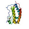
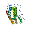
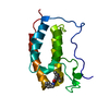
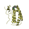
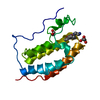
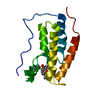
| Title | Crystal Structure of Phospholipase A2 (MIPLA3) From Micropechis Ikaheka | ||||||
 Components Components | PHOSPHOLIPASE A2 | ||||||
 Keywords Keywords | HYDROLASE / PHOSPHOLIPASE A2 / MICROPECHIS IKAHEKA / PANCREATIC LOOP | ||||||
| Function / homology | Phospholipase A2 / Phospholipase A2 domain / Up-down Bundle / Mainly Alpha Function and homology information Function and homology information | ||||||
| Biological species |  Micropechis ikaheka (cobra) Micropechis ikaheka (cobra) | ||||||
| Method |  X-RAY DIFFRACTION / SYNCHROTRON / molecular replacement / Resolution: 2.7 Å X-RAY DIFFRACTION / SYNCHROTRON / molecular replacement / Resolution: 2.7 Å | ||||||
 Authors Authors | Lok, S.M. / Swaminathan, K. | ||||||
 Citation Citation | Journal: FEBS J. / Year: 2005 Title: Structure and function comparison of Micropechis ikaheka snake venom phospholipase A2 isoenzymes Authors: Lok, S.M. / Gao, R. / Rouault, M. / Lambeau, G. / Gopalakrishnakone, P. / Swaminathan, K. #1: Journal: BIOCHIM.BIOPHYS.ACTA / Year: 2001 Title: Purification and properties of three new phospholipase A2 isoenzymes from Micropechis ikaheka venom Authors: Gao, R. / Kini, R.M. / Li, G.D. / Luo, R.H. / Selvanayagam, Z.E. / Gopalakrishnakone, P. | ||||||
| History |
|
- Structure visualization
| Structure viewer | Molecule: MolmilJmol/JSmol |
|---|
- Downloads & links
-Download
| PDBx/mmCIF format | 1ozy.cif.gz | 60.4 KB | Display | PDBx/mmCIF format |
|---|---|---|---|---|
| PDB format | pdb1ozy.ent.gz | 45.3 KB | Display | PDB format |
| PDBx/mmJSON format | 1ozy.json.gz | Tree view | PDBx/mmJSON format | |
| Others |  Other downloads Other downloads |
-Validation report
| Arichive directory | https://data.pdbj.org/pub/pdb/validation_reports/oz/1ozyftp://data.pdbj.org/pub/pdb/validation_reports/oz/1ozy | HTTPS FTP |
|---|
-Related structure data
| Related structure data |  1p7oC  1pwoC  1uneS S: Starting model for refinement C: citing same article ( |
|---|---|
| Similar structure data |


-Links
 PDBj
PDBj
- Assembly
| Deposited unit | 
| ||||||||
|---|---|---|---|---|---|---|---|---|---|
| 1 | 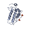
| ||||||||
| 2 | 
| ||||||||
| 3 | 
| ||||||||
| Unit cell |
|
-Components
| #1: Protein | Mass: 13566.533 Da / Num. of mol.: 2 / Source method: isolated from a natural source / Source: (natural) Micropechis ikaheka (cobra) / References: phospholipase A2#2: Chemical |   Mass: 96.063 Da / Num. of mol.: 2 / Source method: obtained synthetically / Formula: SO4 Mass: 96.063 Da / Num. of mol.: 2 / Source method: obtained synthetically / Formula: SO4#3: Water | ChemComp-HOH / |  Mass: 18.015 Da / Num. of mol.: 91 / Source method: isolated from a natural source / Formula: H2O Mass: 18.015 Da / Num. of mol.: 91 / Source method: isolated from a natural source / Formula: H2OHas protein modification | Y | |
|---|
-Experimental details
-Experiment
| Experiment | Method: X-RAY DIFFRACTION / Number of used crystals: 1 |
|---|
- Sample preparation
| Crystal | Density Matthews: 3.83 Å3/Da / Density % sol: 67.59 % |
|---|---|
| Crystal grow | Temperature: 293 K / Method: vapor diffusion, sitting drop / pH: 6.5 Details: ammonium sulphate, pH 6.5, VAPOR DIFFUSION, SITTING DROP, temperature 293K |
-Data collection
| Diffraction | Mean temperature: 100 K |
|---|---|
| Diffraction source | Source: SYNCHROTRON / Site: ALS  / Beamline: 5.0.1 / Wavelength: 0.99 / Wavelength: 0.99 Å / Beamline: 5.0.1 / Wavelength: 0.99 / Wavelength: 0.99 Å |
| Detector | Type: ADSC QUANTUM 4 / Detector: CCD / Date: Jan 27, 2002 |
| Radiation | Monochromator: DIAMOND / Protocol: SINGLE WAVELENGTH / Monochromatic (M) / Laue (L): M / Scattering type: x-ray |
| Radiation wavelength | Wavelength: 0.99 Å / Relative weight: 1 |
| Reflection | Resolution: 2.7→100 Å / Num. obs: 11335 / % possible obs: 98.7 % / Observed criterion σ(I): -3 / Redundancy: 6.8 % / Rsym value: 0.086 / Net I/σ(I): -3 |
| Reflection shell | Resolution: 2.7→2.8 Å / Redundancy: 4.8 % / Rsym value: 0.307 / % possible all: 88.2 |
- Processing
| Software |
| ||||||||||||||||||||||||||||||||||||||||||||||||||||||||||||||||||||||||||||||||
|---|---|---|---|---|---|---|---|---|---|---|---|---|---|---|---|---|---|---|---|---|---|---|---|---|---|---|---|---|---|---|---|---|---|---|---|---|---|---|---|---|---|---|---|---|---|---|---|---|---|---|---|---|---|---|---|---|---|---|---|---|---|---|---|---|---|---|---|---|---|---|---|---|---|---|---|---|---|---|---|---|---|
| Refinement | Method to determine structure: molecular replacement Starting model: PDB CODE: 1UNE Resolution: 2.7→8 Å / Rfactor Rfree error: 0.005 / Data cutoff high absF: 155653.55 / Data cutoff low absF: 0 / Isotropic thermal model: RESTRAINED / Cross valid method: THROUGHOUT / σ(F): 3 Details: TWINNING OPERATOR= h,-k,-l TWINNING FRACTION= 0.474
| ||||||||||||||||||||||||||||||||||||||||||||||||||||||||||||||||||||||||||||||||
| Solvent computation | Solvent model: FLAT MODEL | ||||||||||||||||||||||||||||||||||||||||||||||||||||||||||||||||||||||||||||||||
| Displacement parameters | Biso mean: 51.9541 Å2
| ||||||||||||||||||||||||||||||||||||||||||||||||||||||||||||||||||||||||||||||||
| Refine analyze | Luzzati coordinate error free: 0.47 Å / Luzzati sigma a free: 0.8 Å | ||||||||||||||||||||||||||||||||||||||||||||||||||||||||||||||||||||||||||||||||
| Refinement step | Cycle: LAST / Resolution: 2.7→8 Å
| ||||||||||||||||||||||||||||||||||||||||||||||||||||||||||||||||||||||||||||||||
| Refine LS restraints |
| ||||||||||||||||||||||||||||||||||||||||||||||||||||||||||||||||||||||||||||||||
| LS refinement shell | Resolution: 2.7→2.79 Å / Rfactor Rfree error: 0.005 / Total num. of bins used: 10
| ||||||||||||||||||||||||||||||||||||||||||||||||||||||||||||||||||||||||||||||||
| Xplor file |
|
