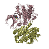
 Movie
Movie Controller


+ Open data

- Basic information
| Entry | Database: PDB / ID: 1oyy | ||||||
|---|---|---|---|---|---|---|---|
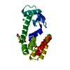
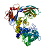
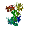
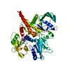
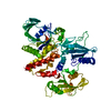
| Title | Structure of the RecQ Catalytic Core bound to ATP-gamma-S | ||||||
 Components Components | ATP-dependent DNA helicase | ||||||
 Keywords Keywords | HYDROLASE / RecQ / helicase / winged helix / helix-turn-helix / ATP binding / Zn(2+) binding / ATP(gamma)S | ||||||
| Function / homology |  Function and homology information Function and homology informationsingle-stranded DNA-dependent ATP-dependent DNA helicase complex / G-quadruplex unwinding activity / bacterial nucleoid / four-way junction helicase activity / SOS response / replisome / single-stranded DNA helicase activity / 3'-5' DNA helicase activity / DNA 3'-5' helicase / transition metal ion binding ...single-stranded DNA-dependent ATP-dependent DNA helicase complex / G-quadruplex unwinding activity / bacterial nucleoid / four-way junction helicase activity / SOS response / replisome / single-stranded DNA helicase activity / 3'-5' DNA helicase activity / DNA 3'-5' helicase / transition metal ion binding / ATP-dependent activity, acting on DNA / DNA helicase activity / chromosome / DNA recombination / DNA replication / DNA repair / DNA damage response / ATP hydrolysis activity / DNA binding / zinc ion binding / ATP binding / cytoplasm Similarity search - Function | ||||||
| Biological species |  | ||||||
| Method |  X-RAY DIFFRACTION / SYNCHROTRON / MOLECULAR REPLACEMENT / Resolution: 2.5 Å X-RAY DIFFRACTION / SYNCHROTRON / MOLECULAR REPLACEMENT / Resolution: 2.5 Å | ||||||
 Authors Authors | Bernstein, D.A. / Zittel, M.C. / Keck, J.L. | ||||||
 Citation Citation | Journal: Embo J. / Year: 2003 Title: High-resolution structure of the E. coli RecQ helicase catalytic core Authors: Bernstein, D.A. / Zittel, M.C. / Keck, J.L. | ||||||
| History |
| ||||||
| Remark 300 | BIOMOLECULE: THIS ENTRY CONTAINS THE CRYSTALLOGRAPHIC ASYMMETRIC UNIT WHICH CONSISTS OF 1 CHAIN(S). ...BIOMOLECULE: THIS ENTRY CONTAINS THE CRYSTALLOGRAPHIC ASYMMETRIC UNIT WHICH CONSISTS OF 1 CHAIN(S). THE AUTHORS STATE IT IS NOT CLEAR WHAT THE ACTIVE OLIGOMERIC STATE OF E. COLI RECQ HELICASE IS AT THIS TIME. |
- Structure visualization
| Structure viewer | Molecule: MolmilJmol/JSmol |
|---|
- Downloads & links
-Download
| PDBx/mmCIF format | 1oyy.cif.gz | 117.8 KB | Display | PDBx/mmCIF format |
|---|---|---|---|---|
| PDB format | pdb1oyy.ent.gz | 88.4 KB | Display | PDB format |
| PDBx/mmJSON format | 1oyy.json.gz | Tree view | PDBx/mmJSON format | |
| Others |  Other downloads Other downloads |
-Validation report
| Arichive directory | https://data.pdbj.org/pub/pdb/validation_reports/oy/1oyyftp://data.pdbj.org/pub/pdb/validation_reports/oy/1oyy | HTTPS FTP |
|---|
-Related structure data
| Related structure data |  1oywSC S: Starting model for refinement C: citing same article ( |
|---|---|
| Similar structure data |


-Links
 PDBj
PDBj


- Assembly
| Deposited unit | 
| ||||||||
|---|---|---|---|---|---|---|---|---|---|
| 1 |
| ||||||||
| Unit cell |
|
-Components
| #1: Protein | Mass: 58730.191 Da / Num. of mol.: 1 / Fragment: 54 kDa Catalytic domain Source method: isolated from a genetically manipulated source Source: (gene. exp.) References: UniProt: P15043, Hydrolases; Acting on acid anhydrides; In phosphorus-containing anhydrides | ||||
|---|---|---|---|---|---|
| #2: Chemical | ChemComp-ZN /   Mass: 65.409 Da / Num. of mol.: 1 / Source method: obtained synthetically / Formula: Zn Mass: 65.409 Da / Num. of mol.: 1 / Source method: obtained synthetically / Formula: Zn | ||||
| #3: Chemical |   Mass: 54.938 Da / Num. of mol.: 2 / Source method: obtained synthetically / Formula: Mn Mass: 54.938 Da / Num. of mol.: 2 / Source method: obtained synthetically / Formula: Mn#4: Chemical | ChemComp-AGS / |   Mass: 523.247 Da / Num. of mol.: 1 / Source method: obtained synthetically / Formula: C10H16N5O12P3S / Comment: ATP-gamma-S, energy-carrying molecule analogue*YM Mass: 523.247 Da / Num. of mol.: 1 / Source method: obtained synthetically / Formula: C10H16N5O12P3S / Comment: ATP-gamma-S, energy-carrying molecule analogue*YM#5: Water | ChemComp-HOH / |  Mass: 18.015 Da / Num. of mol.: 43 / Source method: isolated from a natural source / Formula: H2O Mass: 18.015 Da / Num. of mol.: 43 / Source method: isolated from a natural source / Formula: H2O |
-Experimental details
-Experiment
| Experiment | Method: X-RAY DIFFRACTION / Number of used crystals: 1 |
|---|
- Sample preparation
| Crystal | Density Matthews: 2.27 Å3/Da / Density % sol: 45.9 % | |||||||||||||||||||||||||||||||||||||||||||||||||
|---|---|---|---|---|---|---|---|---|---|---|---|---|---|---|---|---|---|---|---|---|---|---|---|---|---|---|---|---|---|---|---|---|---|---|---|---|---|---|---|---|---|---|---|---|---|---|---|---|---|---|
| Crystal grow | Method: vapor diffusion, hanging drop / pH: 7.5 Details: PEG 1000, PEG 400, MES, Ammonium Sulfate, Manganese Chloride, pH 7.5, VAPOR DIFFUSION, HANGING DROP, temperature 110K | |||||||||||||||||||||||||||||||||||||||||||||||||
| Crystal grow | *PLUS pH: 6.5 / Method: vapor diffusion, hanging drop | |||||||||||||||||||||||||||||||||||||||||||||||||
| Components of the solutions | *PLUS
|
-Data collection
| Diffraction | Mean temperature: 110 K |
|---|---|
| Diffraction source | Source: SYNCHROTRON / Site: APS  / Beamline: 14-BM-C / Wavelength: 0.9 Å / Beamline: 14-BM-C / Wavelength: 0.9 Å |
| Detector | Type: ADSC QUANTUM 4 / Detector: CCD / Date: Feb 16, 2003 |
| Radiation | Protocol: SINGLE WAVELENGTH / Monochromatic (M) / Laue (L): M / Scattering type: x-ray |
| Radiation wavelength | Wavelength: 0.9 Å / Relative weight: 1 |
| Reflection | Resolution: 2.5→20 Å / Num. all: 18473 / Num. obs: 16814 / % possible obs: 91 % / Observed criterion σ(F): 0 / Observed criterion σ(I): 0 / Redundancy: 2.7 % / Rsym value: 0.048 / Net I/σ(I): 12.8 |
| Reflection shell | Resolution: 2.5→2.63 Å / Mean I/σ(I) obs: 7.1 / Rsym value: 0.133 / % possible all: 92.1 |
| Reflection | *PLUS Lowest resolution: 21 Å / Num. obs: 17529 / % possible obs: 92.1 % / Num. measured all: 47206 / Rmerge(I) obs: 0.048 |
| Reflection shell | *PLUS % possible obs: 92.1 % / Rmerge(I) obs: 0.133 |
- Processing
| Software |
| ||||||||||||||||||||
|---|---|---|---|---|---|---|---|---|---|---|---|---|---|---|---|---|---|---|---|---|---|
| Refinement | Method to determine structure: MOLECULAR REPLACEMENT Starting model: PDB entry 1OYW Resolution: 2.5→20 Å / σ(F): 0 / Stereochemistry target values: Engh & Huber
| ||||||||||||||||||||
| Refinement step | Cycle: LAST / Resolution: 2.5→20 Å
| ||||||||||||||||||||
| Refine LS restraints |
| ||||||||||||||||||||
| LS refinement shell | Resolution: 2.5→2.614 Å
| ||||||||||||||||||||
| Refinement | *PLUS Lowest resolution: 20 Å / % reflection Rfree: 5 % / Rfactor Rwork: 0.21 | ||||||||||||||||||||
| Solvent computation | *PLUS | ||||||||||||||||||||
| Displacement parameters | *PLUS | ||||||||||||||||||||
| Refine LS restraints | *PLUS
|
