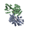
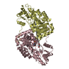
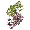
 Movie
Movie Controller


+ Open data

- Basic information
| Entry | Database: PDB / ID: 1oxo | ||||||
|---|---|---|---|---|---|---|---|
| Title | ASPARTATE AMINOTRANSFERASE, H-ASP COMPLEX, OPEN CONFORMATION | ||||||
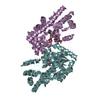
 Components Components | ASPARTATE AMINOTRANSFERASE | ||||||
 Keywords Keywords | AMINOTRANSFERASE / VITAMIN B6 / HYDROXYLAMINE DERIVED INHIBITORS | ||||||
| Function / homology |  Function and homology information Function and homology informationAmino acid metabolism / Gluconeogenesis / L-aspartate catabolic process / kynurenine-oxoglutarate transaminase / L-kynurenine:2-oxoglutarate transaminase activity / aspartate metabolic process / glutamate metabolic process / aspartate transaminase / L-aspartate:2-oxoglutarate transaminase activity / 2-oxoglutarate metabolic process ...Amino acid metabolism / Gluconeogenesis / L-aspartate catabolic process / kynurenine-oxoglutarate transaminase / L-kynurenine:2-oxoglutarate transaminase activity / aspartate metabolic process / glutamate metabolic process / aspartate transaminase / L-aspartate:2-oxoglutarate transaminase activity / 2-oxoglutarate metabolic process / pyridoxal phosphate binding / mitochondrial matrix / protein homodimerization activity / mitochondrion Similarity search - Function | ||||||
| Biological species |  | ||||||
| Method |  X-RAY DIFFRACTION / Resolution: 2.3 Å X-RAY DIFFRACTION / Resolution: 2.3 Å | ||||||
 Authors Authors | Hohenester, E. / Schirmer, T. / Jansonius, J.N. | ||||||
 Citation Citation | Journal: Eur.J.Biochem. / Year: 1996 Title: Crystal structures and solution studies of oxime adducts of mitochondrial aspartate aminotransferase. Authors: Markovic-Housley, Z. / Schirmer, T. / Hohenester, E. / Khomutov, A.R. / Khomutov, R.M. / Karpeisky, M.Y. / Sandmeier, E. / Christen, P. / Jansonius, J.N. #1: Journal: J.Mol.Biol. / Year: 1992Title: X-Ray Structure Refinement and Comparison of Three Forms of Mitochondrial Aspartate Aminotransferase Authors: Mcphalen, C.A. / Vincent, M.G. / Jansonius, J.N. | ||||||
| History |
|
- Structure visualization
| Structure viewer | Molecule: MolmilJmol/JSmol |
|---|
- Downloads & links
-Download
| PDBx/mmCIF format | 1oxo.cif.gz | 180.6 KB | Display | PDBx/mmCIF format |
|---|---|---|---|---|
| PDB format | pdb1oxo.ent.gz | 141.2 KB | Display | PDB format |
| PDBx/mmJSON format | 1oxo.json.gz | Tree view | PDBx/mmJSON format | |
| Others |  Other downloads Other downloads |
-Validation report
| Arichive directory | https://data.pdbj.org/pub/pdb/validation_reports/ox/1oxoftp://data.pdbj.org/pub/pdb/validation_reports/ox/1oxo | HTTPS FTP |
|---|
-Related structure data


-Links
 PDBj
PDBj- Assembly
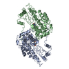
| Deposited unit | 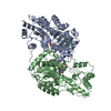
| ||||||||
|---|---|---|---|---|---|---|---|---|---|
| 1 |
| ||||||||
| Unit cell |
| ||||||||
| Noncrystallographic symmetry (NCS) | NCS oper: (Code: given Matrix: (-0.92544, 0.34253, -0.16199), Vector: Details | THE MOLECULE IS AN ALPHA2 DIMER. THE SUBUNITS ARE RELATED BY A LOCAL TWO-FOLD ROTATION AXIS. THEY ARE DISTINGUISHED BY CHAIN IDENTIFIERS A AND B, RESPECTIVELY. THE RESIDUES IN EACH SUBUNIT ARE NUMBERED FROM 3 TO 410, ACCORDING TO A SEQUENCE ALIGNMENT WITH THE LONGER SEQUENCE OF PIG CYTOSOLIC ASPARTATE AMINOTRANSFERASE (SEE U. GRAF-HAUSNER, K.J. WILSON AND P. CHRISTEN (1983) J.BIOL.CHEM. 258, 8813-8826). | |
-Components
| #1: Protein | Mass: 44992.484 Da / Num. of mol.: 2 Source method: isolated from a genetically manipulated source Source: (gene. exp.) #2: Chemical | 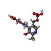  Mass: 322.208 Da / Num. of mol.: 2 / Source method: obtained synthetically / Formula: C10H15N2O8P Mass: 322.208 Da / Num. of mol.: 2 / Source method: obtained synthetically / Formula: C10H15N2O8P#3: Water | ChemComp-HOH / |  Mass: 18.015 Da / Num. of mol.: 541 / Source method: isolated from a natural source / Formula: H2O Mass: 18.015 Da / Num. of mol.: 541 / Source method: isolated from a natural source / Formula: H2O |
|---|
-Experimental details
-Experiment
| Experiment | Method: X-RAY DIFFRACTION |
|---|
- Sample preparation
| Crystal | Density Matthews: 2.35 Å3/Da / Density % sol: 47.6 % Description: BECAUSE OF THE LOW REDUNDANCY, THE DATA WERE SCALED TO A REFERENCE DATA SET CALCULATED FROM THE COORDINATES OF THE HOLO-AAT STRUCTURE REFINED AT 1.9 ANGSTROMS RESOLUTION (MCPHALEN ET AL., 1992). | ||||||||||||||||||||||||||||||
|---|---|---|---|---|---|---|---|---|---|---|---|---|---|---|---|---|---|---|---|---|---|---|---|---|---|---|---|---|---|---|---|
| Crystal grow | *PLUS pH: 7.5 / Method: vapor diffusion, hanging drop | ||||||||||||||||||||||||||||||
| Components of the solutions | *PLUS
|
-Data collection
| Diffraction source | Wavelength: 1.5418 |
|---|---|
| Detector | Type: ENRAF-NONIUS FAST / Detector: DIFFRACTOMETER / Date: Jan 1, 1991 |
| Radiation | Monochromatic (M) / Laue (L): M / Scattering type: x-ray |
| Radiation wavelength | Wavelength: 1.5418 Å / Relative weight: 1 |
| Reflection | Num. obs: 25752 / Redundancy: 1.21 % / Rmerge(I) obs: 0.041 |
- Processing
| Software |
| ||||||||||||||||||||||||||||||||||||||||||||||||||||||||||||||||||||||||||||||||||||
|---|---|---|---|---|---|---|---|---|---|---|---|---|---|---|---|---|---|---|---|---|---|---|---|---|---|---|---|---|---|---|---|---|---|---|---|---|---|---|---|---|---|---|---|---|---|---|---|---|---|---|---|---|---|---|---|---|---|---|---|---|---|---|---|---|---|---|---|---|---|---|---|---|---|---|---|---|---|---|---|---|---|---|---|---|---|
| Refinement | Resolution: 2.3→8 Å / Num. reflection obs: 25169 / σ(F): 0 | ||||||||||||||||||||||||||||||||||||||||||||||||||||||||||||||||||||||||||||||||||||
| Displacement parameters | Biso mean: 18.7 Å2 | ||||||||||||||||||||||||||||||||||||||||||||||||||||||||||||||||||||||||||||||||||||
| Refinement step | Cycle: LAST / Resolution: 2.3→8 Å
| ||||||||||||||||||||||||||||||||||||||||||||||||||||||||||||||||||||||||||||||||||||
| Refine LS restraints |
| ||||||||||||||||||||||||||||||||||||||||||||||||||||||||||||||||||||||||||||||||||||
| Software | *PLUS Name: PROLSQ / Classification: refinement | ||||||||||||||||||||||||||||||||||||||||||||||||||||||||||||||||||||||||||||||||||||
| Refinement | *PLUS Rfactor Rwork: 0.128 | ||||||||||||||||||||||||||||||||||||||||||||||||||||||||||||||||||||||||||||||||||||
| Solvent computation | *PLUS | ||||||||||||||||||||||||||||||||||||||||||||||||||||||||||||||||||||||||||||||||||||
| Displacement parameters | *PLUS |
