 Movie
Movie Controller


+ Open data

- Basic information
| Entry | Database: PDB / ID: 1ox9 | ||||||
|---|---|---|---|---|---|---|---|
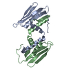
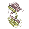
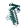
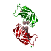
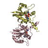
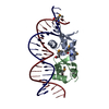
| Title | Crystal structure of SspB-ssrA complex | ||||||
 Components Components |
| ||||||
 Keywords Keywords | HYDROLASE ACTIVATOR / SspB-ssrA | ||||||
| Function / homology |  Function and homology information Function and homology information: / HslUV protease complex / positive regulation of ATP-dependent activity / protein catabolic process / positive regulation of protein catabolic process / ATPase binding / molecular adaptor activity / ribosome / protein homodimerization activity / RNA binding / cytosol Similarity search - Function | ||||||
| Biological species |  | ||||||
| Method |  X-RAY DIFFRACTION / SYNCHROTRON / MOLECULAR REPLACEMENT / Resolution: 2.9 Å X-RAY DIFFRACTION / SYNCHROTRON / MOLECULAR REPLACEMENT / Resolution: 2.9 Å | ||||||
 Authors Authors | Song, H.K. / Eck, M.J. | ||||||
 Citation Citation | Journal: Mol.Cell / Year: 2003 Title: Structural basis of degradation signal recognition by SspB, a specificity-enhancing factor for the ClpXP proteolytic machine Authors: Song, H.K. / Eck, M.J. | ||||||
| History |
|
- Structure visualization
| Structure viewer | Molecule: MolmilJmol/JSmol |
|---|
- Downloads & links
-Download
| PDBx/mmCIF format | 1ox9.cif.gz | 180.1 KB | Display | PDBx/mmCIF format |
|---|---|---|---|---|
| PDB format | pdb1ox9.ent.gz | 148.2 KB | Display | PDB format |
| PDBx/mmJSON format | 1ox9.json.gz | Tree view | PDBx/mmJSON format | |
| Others |  Other downloads Other downloads |
-Validation report
| Arichive directory | https://data.pdbj.org/pub/pdb/validation_reports/ox/1ox9ftp://data.pdbj.org/pub/pdb/validation_reports/ox/1ox9 | HTTPS FTP |
|---|
-Related structure data


-Links
 PDBj
PDBj- Assembly
| Deposited unit | 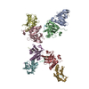
| ||||||||
|---|---|---|---|---|---|---|---|---|---|
| 1 | 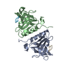
| ||||||||
| 2 | 
| ||||||||
| 3 | 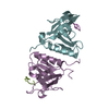
| ||||||||
| 4 | 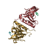
| ||||||||
| Unit cell |
|
-Components
| #1: Protein | Mass: 12018.668 Da / Num. of mol.: 8 / Fragment: Residues 4-111 Source method: isolated from a genetically manipulated source Source: (gene. exp.) #2: Protein/peptide | Mass: 866.830 Da / Num. of mol.: 8 / Source method: obtained synthetically Details: This ssrA peptide is synthesized and occurs naturally in E. coli. |
|---|
-Experimental details
-Experiment
| Experiment | Method: X-RAY DIFFRACTION / Number of used crystals: 1 |
|---|
- Sample preparation
| Crystal | Density Matthews: 4.29 Å3/Da / Density % sol: 71.11 % | |||||||||||||||||||||||||||||||||||||||||||||||||||||||||||||||
|---|---|---|---|---|---|---|---|---|---|---|---|---|---|---|---|---|---|---|---|---|---|---|---|---|---|---|---|---|---|---|---|---|---|---|---|---|---|---|---|---|---|---|---|---|---|---|---|---|---|---|---|---|---|---|---|---|---|---|---|---|---|---|---|---|
| Crystal grow | Temperature: 295 K / Method: vapor diffusion, hanging drop / pH: 8 Details: PEG 8K, pH 8.0, VAPOR DIFFUSION, HANGING DROP, temperature 295K | |||||||||||||||||||||||||||||||||||||||||||||||||||||||||||||||
| Crystal grow | *PLUS Temperature: 22 ℃ / pH: 7.7 / Method: vapor diffusion, hanging drop | |||||||||||||||||||||||||||||||||||||||||||||||||||||||||||||||
| Components of the solutions | *PLUS
|
-Data collection
| Diffraction | Mean temperature: 100 K |
|---|---|
| Diffraction source | Source: SYNCHROTRON / Site: CHESS  / Beamline: A1 / Wavelength: 0.95 Å / Beamline: A1 / Wavelength: 0.95 Å |
| Detector | Type: ADSC QUANTUM 4 / Detector: CCD / Date: Nov 30, 2001 |
| Radiation | Monochromator: Si 111 CHANNEL / Protocol: SINGLE WAVELENGTH / Monochromatic (M) / Laue (L): M / Scattering type: x-ray |
| Radiation wavelength | Wavelength: 0.95 Å / Relative weight: 1 |
| Reflection | Resolution: 2.9→30 Å / Num. obs: 34398 / % possible obs: 93.4 % / Observed criterion σ(F): 3 / Observed criterion σ(I): 3 |
| Reflection shell | Resolution: 2.9→2.99 Å / % possible all: 89.9 |
| Reflection | *PLUS Num. measured all: 86232 / Rmerge(I) obs: 0.124 |
| Reflection shell | *PLUS % possible obs: 89.9 % / Rmerge(I) obs: 0.236 |
- Processing
| Software |
| ||||||||||||||||||||
|---|---|---|---|---|---|---|---|---|---|---|---|---|---|---|---|---|---|---|---|---|---|
| Refinement | Method to determine structure: MOLECULAR REPLACEMENT / Resolution: 2.9→30 Å / σ(F): 0 / Stereochemistry target values: Engh & Huber
| ||||||||||||||||||||
| Refinement step | Cycle: LAST / Resolution: 2.9→30 Å
| ||||||||||||||||||||
| Refine LS restraints |
| ||||||||||||||||||||
| Refinement | *PLUS Lowest resolution: 30 Å / % reflection Rfree: 10 % / Rfactor Rwork: 0.245 | ||||||||||||||||||||
| Solvent computation | *PLUS | ||||||||||||||||||||
| Displacement parameters | *PLUS |
