 Movie
Movie Controller

[English] 日本語
 Yorodumi
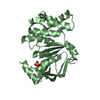
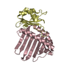
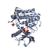
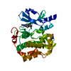
- PDB-1ox7: Crystal structure of yeast cytosine deaminase apo-enzyme: inorgan... -
+ Open data

- Basic information
| Entry | Database: PDB / ID: 1ox7 | ||||||
|---|---|---|---|---|---|---|---|
| Title | Crystal structure of yeast cytosine deaminase apo-enzyme: inorganic zinc bound | ||||||
 Components Components | Cytosine deaminase | ||||||
 Keywords Keywords | HYDROLASE / aminohydrolase | ||||||
| Function / homology |  Function and homology information Function and homology informationcytidine metabolic process / pyrimidine-containing compound salvage / diaminohydroxyphosphoribosylaminopyrimidine deaminase activity / cytosine deaminase / cytosine deaminase activity / UMP salvage / cytosine metabolic process / zinc ion binding / nucleus / cytoplasm Similarity search - Function | ||||||
| Biological species |  | ||||||
| Method |  X-RAY DIFFRACTION / SYNCHROTRON / SAD / Resolution: 1.43 Å X-RAY DIFFRACTION / SYNCHROTRON / SAD / Resolution: 1.43 Å | ||||||
 Authors Authors | Ireton, G.C. / Black, M.E. / Stoddard, B.L. | ||||||
 Citation Citation | Journal: Structure / Year: 2003 Title: The 1.14 a crystal structure of yeast Cytosine deaminase. Evolution of nucleotide salvage enzymes and implications for genetic chemotherapy. Authors: Ireton, G.C. / Black, M.E. / Stoddard, B.L. | ||||||
| History |
|
- Structure visualization
| Structure viewer | Molecule: MolmilJmol/JSmol |
|---|
- Downloads & links
-Download
| PDBx/mmCIF format | 1ox7.cif.gz | 84 KB | Display | PDBx/mmCIF format |
|---|---|---|---|---|
| PDB format | pdb1ox7.ent.gz | 62 KB | Display | PDB format |
| PDBx/mmJSON format | 1ox7.json.gz | Tree view | PDBx/mmJSON format | |
| Others |  Other downloads Other downloads |
-Validation report
| Arichive directory | https://data.pdbj.org/pub/pdb/validation_reports/ox/1ox7ftp://data.pdbj.org/pub/pdb/validation_reports/ox/1ox7 | HTTPS FTP |
|---|
-Related structure data


-Links
 PDBj
PDBj- Assembly
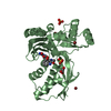
| Deposited unit | 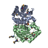
| ||||||||
|---|---|---|---|---|---|---|---|---|---|
| 1 |
| ||||||||
| Unit cell |
|
-Components
| #1: Protein | Mass: 18089.492 Da / Num. of mol.: 2 Source method: isolated from a genetically manipulated source Source: (gene. exp.) Gene: FCY1 OR YPR062W OR YP9499.17 / Plasmid: pET15b-(yCD) / Production host:  #2: Chemical | ChemComp-ZN /   Mass: 65.409 Da / Num. of mol.: 4 / Source method: obtained synthetically / Formula: Zn Mass: 65.409 Da / Num. of mol.: 4 / Source method: obtained synthetically / Formula: Zn#3: Chemical | ChemComp-CA / |   Mass: 40.078 Da / Num. of mol.: 1 / Source method: obtained synthetically / Formula: Ca Mass: 40.078 Da / Num. of mol.: 1 / Source method: obtained synthetically / Formula: Ca#4: Water | ChemComp-HOH / |  Mass: 18.015 Da / Num. of mol.: 375 / Source method: isolated from a natural source / Formula: H2O Mass: 18.015 Da / Num. of mol.: 375 / Source method: isolated from a natural source / Formula: H2OHas protein modification | Y | |
|---|
-Experimental details
-Experiment
| Experiment | Method: X-RAY DIFFRACTION / Number of used crystals: 1 |
|---|
- Sample preparation
| Crystal | Density Matthews: 1.74 Å3/Da / Density % sol: 28.68 % | ||||||||||||||||||||||||||||||||||||||||
|---|---|---|---|---|---|---|---|---|---|---|---|---|---|---|---|---|---|---|---|---|---|---|---|---|---|---|---|---|---|---|---|---|---|---|---|---|---|---|---|---|---|
| Crystal grow | Temperature: 277 K / Method: streak seeding / pH: 6.5 Details: PEG 8000, calcium acetate, sodium cacodylate, pH 6.5, streak seeding, temperature 277K | ||||||||||||||||||||||||||||||||||||||||
| Crystal grow | *PLUS Temperature: 4. ℃ / Method: vapor diffusion, hanging drop | ||||||||||||||||||||||||||||||||||||||||
| Components of the solutions | *PLUS
|
-Data collection
| Diffraction | Mean temperature: 100 K |
|---|---|
| Diffraction source | Source: SYNCHROTRON / Site: ALS  / Beamline: 5.0.2 / Wavelength: 0.9795 Å / Beamline: 5.0.2 / Wavelength: 0.9795 Å |
| Detector | Type: ADSC QUANTUM 210 / Detector: CCD / Date: Sep 16, 2002 |
| Radiation | Monochromator: double crystal Si(111) / Protocol: MAD / Monochromatic (M) / Laue (L): M / Scattering type: x-ray |
| Radiation wavelength | Wavelength: 0.9795 Å / Relative weight: 1 |
| Reflection | Resolution: 1.43→20 Å / Num. all: 98785 / Num. obs: 98785 / % possible obs: 99.9 % / Observed criterion σ(F): 0 / Observed criterion σ(I): 0 / Redundancy: 7.4 % / Biso Wilson estimate: 9.6 Å2 / Rmerge(I) obs: 0.048 / Net I/σ(I): 27.4 |
| Reflection shell | Resolution: 1.43→1.48 Å / Rmerge(I) obs: 0.165 / Mean I/σ(I) obs: 9.5 / Num. unique all: 9823 / % possible all: 99.7 |
| Reflection | *PLUS Rmerge(I) obs: 0.042 |
| Reflection shell | *PLUS % possible obs: 99.7 % |
- Processing
| Software |
| ||||||||||||||||||||||||||||||||||||
|---|---|---|---|---|---|---|---|---|---|---|---|---|---|---|---|---|---|---|---|---|---|---|---|---|---|---|---|---|---|---|---|---|---|---|---|---|---|
| Refinement | Method to determine structure: SAD / Resolution: 1.43→19.83 Å / Rfactor Rfree error: 0.003 / Isotropic thermal model: RESTRAINED / σ(F): 0 / Stereochemistry target values: Engh & Huber
| ||||||||||||||||||||||||||||||||||||
| Solvent computation | Bsol: 49.5037 Å2 / ksol: 0.376664 e/Å3 | ||||||||||||||||||||||||||||||||||||
| Displacement parameters | Biso mean: 10.8 Å2
| ||||||||||||||||||||||||||||||||||||
| Refine analyze | Luzzati coordinate error free: 0.14 Å / Luzzati sigma a free: 0.07 Å | ||||||||||||||||||||||||||||||||||||
| Refinement step | Cycle: LAST / Resolution: 1.43→19.83 Å
| ||||||||||||||||||||||||||||||||||||
| Refine LS restraints |
| ||||||||||||||||||||||||||||||||||||
| LS refinement shell | Resolution: 1.43→1.52 Å / Rfactor Rfree error: 0.007 / Total num. of bins used: 6
| ||||||||||||||||||||||||||||||||||||
| Xplor file |
| ||||||||||||||||||||||||||||||||||||
| Refinement | *PLUS Lowest resolution: 20 Å / Num. reflection obs: 51872 / Num. reflection Rfree: 2594 / % reflection Rfree: 5 % | ||||||||||||||||||||||||||||||||||||
| Solvent computation | *PLUS | ||||||||||||||||||||||||||||||||||||
| Displacement parameters | *PLUS | ||||||||||||||||||||||||||||||||||||
| Refine LS restraints | *PLUS
|
