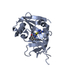
[English] 日本語
Yorodumi
- PDB-1oh4: Structural and thermodynamic dissection of specific mannan recogn... -

+
Open data


ID or keywords:

Loading...

-
Basic information

Entry
Database: PDB / ID: 1oh4
TitleStructural and thermodynamic dissection of specific mannan recognition by a carbohydrate-binding module
ComponentsBETA-MANNOSIDASE
KeywordsHYDROLASE / CARBOHYDRATE BINDING MODULE / MANNAN / LECTIN / GLYCOSIDE HYDROLASE
Function / homology
Function and homology information


organic substance metabolic process / hydrolase activity, hydrolyzing O-glycosyl compounds / extracellular region / metal ion binding
Similarity search - Function
Carbohydrate binding module 27 / Carbohydrate binding module 27 / Mannan endo-1,4-beta-mannosidase-like / Glycoside hydrolase, family 5 / Cellulase (glycosyl hydrolase family 5) / Galactose-binding domain-like / Galactose-binding-like domain superfamily / Glycoside hydrolase superfamily / Jelly Rolls / Sandwich / Mainly Beta
Similarity search - Domain/homology
Biological speciesTHERMOTOGA MARITIMA (bacteria)
MethodX-RAY DIFFRACTION / SYNCHROTRON / MOLECULAR REPLACEMENT / Resolution: 1.35 Å
AuthorsBoraston, A.B. / Revett, T.J. / Boraston, C.M. / Nurizzo, D. / Davies, G.J.
CitationJournal: Structure / Year: 2003
Title: Structural and Thermodynamic Dissection of Specific Mannan Recognition by a Carbohydrate Binding Module, Tmcbm27
Authors: Boraston, A.B. / Revett, T.J. / Boraston, C.M. / Nurizzo, D. / Davies, G.J.
History
DepositionMay 21, 2003Deposition site: PDBE / Processing site: PDBE
Revision 1.0Mar 16, 2004Provider: repository / Type: Initial release
Revision 1.1May 8, 2011Group: Version format compliance
Revision 1.2Jul 13, 2011Group: Version format compliance
Revision 2.0Jul 29, 2020Group: Advisory / Atomic model ...Advisory / Atomic model / Data collection / Derived calculations / Other / Structure summary
Category: atom_site / atom_site_anisotrop ...atom_site / atom_site_anisotrop / chem_comp / entity / pdbx_branch_scheme / pdbx_chem_comp_identifier / pdbx_database_status / pdbx_entity_branch / pdbx_entity_branch_descriptor / pdbx_entity_branch_link / pdbx_entity_branch_list / pdbx_entity_nonpoly / pdbx_nonpoly_scheme / pdbx_struct_assembly_gen / pdbx_struct_conn_angle / pdbx_unobs_or_zero_occ_atoms / pdbx_validate_close_contact / struct_asym / struct_conn / struct_site / struct_site_gen
Item: _atom_site.B_iso_or_equiv / _atom_site.Cartn_x ..._atom_site.B_iso_or_equiv / _atom_site.Cartn_x / _atom_site.Cartn_y / _atom_site.Cartn_z / _atom_site.auth_asym_id / _atom_site.auth_seq_id / _atom_site.label_asym_id / _atom_site.label_entity_id / _atom_site_anisotrop.U[1][1] / _atom_site_anisotrop.U[1][2] / _atom_site_anisotrop.U[1][3] / _atom_site_anisotrop.U[2][2] / _atom_site_anisotrop.U[2][3] / _atom_site_anisotrop.U[3][3] / _atom_site_anisotrop.pdbx_auth_asym_id / _atom_site_anisotrop.pdbx_auth_seq_id / _atom_site_anisotrop.pdbx_label_asym_id / _chem_comp.name / _chem_comp.type / _pdbx_database_status.status_code_sf / _pdbx_struct_assembly_gen.asym_id_list / _pdbx_struct_conn_angle.ptnr1_auth_comp_id / _pdbx_struct_conn_angle.ptnr1_auth_seq_id / _pdbx_struct_conn_angle.ptnr1_label_asym_id / _pdbx_struct_conn_angle.ptnr1_label_atom_id / _pdbx_struct_conn_angle.ptnr1_label_comp_id / _pdbx_struct_conn_angle.ptnr1_label_seq_id / _pdbx_struct_conn_angle.ptnr2_label_asym_id / _pdbx_struct_conn_angle.ptnr3_auth_comp_id / _pdbx_struct_conn_angle.ptnr3_auth_seq_id / _pdbx_struct_conn_angle.ptnr3_label_asym_id / _pdbx_struct_conn_angle.ptnr3_label_atom_id / _pdbx_struct_conn_angle.ptnr3_label_comp_id / _pdbx_struct_conn_angle.ptnr3_label_seq_id / _pdbx_struct_conn_angle.value / _pdbx_validate_close_contact.auth_asym_id_1 / _pdbx_validate_close_contact.auth_asym_id_2 / _pdbx_validate_close_contact.auth_seq_id_1 / _pdbx_validate_close_contact.auth_seq_id_2 / _struct_conn.pdbx_dist_value / _struct_conn.pdbx_leaving_atom_flag / _struct_conn.ptnr1_auth_asym_id / _struct_conn.ptnr1_auth_comp_id / _struct_conn.ptnr1_auth_seq_id / _struct_conn.ptnr1_label_asym_id / _struct_conn.ptnr1_label_atom_id / _struct_conn.ptnr1_label_comp_id / _struct_conn.ptnr1_label_seq_id / _struct_conn.ptnr2_auth_asym_id / _struct_conn.ptnr2_auth_comp_id / _struct_conn.ptnr2_auth_seq_id / _struct_conn.ptnr2_label_asym_id / _struct_conn.ptnr2_label_atom_id / _struct_conn.ptnr2_label_comp_id / _struct_conn.ptnr2_label_seq_id
Description: Carbohydrate remediation / Provider: repository / Type: Remediation
Revision 2.1Dec 13, 2023Group: Advisory / Data collection ...Advisory / Data collection / Database references / Refinement description / Structure summary
Category: chem_comp / chem_comp_atom ...chem_comp / chem_comp_atom / chem_comp_bond / database_2 / pdbx_initial_refinement_model / pdbx_unobs_or_zero_occ_atoms
Item: _chem_comp.pdbx_synonyms / _database_2.pdbx_DOI / _database_2.pdbx_database_accession
Remark 700 SHEET THE SHEET STRUCTURE OF THIS MOLECULE IS BIFURCATED. IN ORDER TO REPRESENT THIS FEATURE IN ... SHEET THE SHEET STRUCTURE OF THIS MOLECULE IS BIFURCATED. IN ORDER TO REPRESENT THIS FEATURE IN THE SHEET RECORDS BELOW, TWO SHEETS ARE DEFINED.

-
Structure visualization

Structure viewerMolecule:
MolmilJmol/JSmol

Downloads & links

-
Assembly

Deposited unit
A: BETA-MANNOSIDASE
hetero molecules


Theoretical massNumber of molelcules
Total (without water)21,9626
Polymers20,4891
Non-polymers1,4735
Water3,279182
1


  • Idetical with deposited unit
  • defined by author&software
TypeNameSymmetry operationNumber
identity operation1_555x,y,z1
MethodPQS
Unit cell
Length a, b, c (Å)31.790, 48.919, 96.421
Angle α, β, γ (deg.)90.00, 90.00, 90.00
Int Tables number19
Space group name H-MP212121

-
Components

-
Protein / Sugars , 2 types, 2 molecules A

#1: Protein BETA-MANNOSIDASE / / MANB / TMCBM27


Mass: 20489.016 Da / Num. of mol.: 1 / Fragment: CARBOHYDRATE-BINDING MODULE, RESIDUES 505-680
Source method: isolated from a genetically manipulated source
Source: (gene. exp.) THERMOTOGA MARITIMA (bacteria) / Strain: MSB8 / Plasmid: PET 28 TMCBM27 / Production host: ESCHERICHIA COLI (E. coli) / Strain (production host): BL21(DE3) / References: UniProt: Q9RIK9
#2: Polysaccharide beta-D-mannopyranose-(1-4)-[alpha-D-galactopyranose-(1-6)]beta-D-mannopyranose-(1-4)-[alpha-D- ...beta-D-mannopyranose-(1-4)-[alpha-D-galactopyranose-(1-6)]beta-D-mannopyranose-(1-4)-[alpha-D-galactopyranose-(1-6)]beta-D-mannopyranose-(1-4)-beta-D-mannopyranose-(1-4)-beta-D-mannopyranose


Type: oligosaccharide / Mass: 1153.001 Da / Num. of mol.: 1
Source method: isolated from a genetically manipulated source
DescriptorTypeProgram
DManpb1-4[DGalpa1-6]DManpb1-4[DGalpa1-6]DManpb1-4DManpb1-4DManpb1-ROHGlycam Condensed SequenceGMML 1.0
WURCS=2.0/2,7,6/[a1122h-1b_1-5][a2112h-1a_1-5]/1-1-1-1-1-2-2/a4-b1_b4-c1_c4-d1_c6-g1_d4-e1_d6-f1WURCSPDB2Glycan 1.1.0
[][b-D-Manp]{[(4+1)][b-D-Manp]{[(4+1)][b-D-Manp]{[(4+1)][b-D-Manp]{[(4+1)][b-D-Manp]{}[(6+1)][a-D-Galp]{}}[(6+1)][a-D-Galp]{}}}}LINUCSPDB-CARE

-
Non-polymers , 4 types, 186 molecules

#3: Chemical ChemComp-GOL / GLYCEROL / GLYCERIN / PROPANE-1,2,3-TRIOL / Glycerol


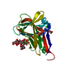
Mass: 92.094 Da / Num. of mol.: 2 / Source method: obtained synthetically / Formula: C3H8O3
#4: Chemical ChemComp-CA / CALCIUM ION


Mass: 40.078 Da / Num. of mol.: 1 / Source method: obtained synthetically / Formula: Ca
#5: Chemical ChemComp-SO4 / SULFATE ION / Sulfate


Mass: 96.063 Da / Num. of mol.: 1 / Source method: obtained synthetically / Formula: SO4
#6: Water ChemComp-HOH / water / Water


Mass: 18.015 Da / Num. of mol.: 182 / Source method: isolated from a natural source / Formula: H2O

-
Experimental details

-
Experiment

ExperimentMethod: X-RAY DIFFRACTION / Number of used crystals: 1

-
Sample preparation

CrystalDensity Matthews: 1.83 Å3/Da / Density % sol: 32 %
Crystal growpH: 4.6
Details: 1.5 M AMMONIUM SULFATE, 0.1 M SODIUM ACETATE PH 4.6

-
Data collection

DiffractionMean temperature: 100 K
Diffraction sourceSource: SYNCHROTRON / Site: ESRF / Beamline: ID14-1 / Wavelength: 0.934
DetectorType: ADSC CCD / Detector: CCD
RadiationProtocol: SINGLE WAVELENGTH / Monochromatic (M) / Laue (L): M / Scattering type: x-ray
Radiation wavelengthWavelength: 0.934 Å / Relative weight: 1
ReflectionResolution: 1.35→1.42 Å / Num. obs: 31455 / % possible obs: 97.8 % / Redundancy: 4.7 % / Rmerge(I) obs: 0.057 / Net I/σ(I): 17.5
Reflection shellResolution: 1.35→1.42 Å / Redundancy: 2.5 % / Rmerge(I) obs: 0.261 / Mean I/σ(I) obs: 2.9 / % possible all: 87.3

-
Processing

Software
NameVersionClassification
REFMAC5.1.24refinement
MOSFLMdata reduction
SCALEPACKdata scaling
AMoREphasing
RefinementMethod to determine structure: MOLECULAR REPLACEMENT
Starting model: PDB ENTRY 1OF3
Resolution: 1.35→48.22 Å / Cor.coef. Fo:Fc: 0.973 / Cor.coef. Fo:Fc free: 0.956 / SU B: 0.721 / SU ML: 0.03 / Cross valid method: THROUGHOUT / ESU R: 0.062 / ESU R Free: 0.055 / Stereochemistry target values: MAXIMUM LIKELIHOOD / Details: HYDROGENS HAVE BEEN ADDED IN THE RIDING POSITIONS
RfactorNum. reflection% reflectionSelection details
Rfree0.17 1680 5.1 %RANDOM
Rwork0.138 ---
obs0.14 31455 97.7 %-
Solvent computationIon probe radii: 0.8 Å / Shrinkage radii: 0.8 Å / VDW probe radii: 1.4 Å / Solvent model: BABINET MODEL WITH MASK
Displacement parametersBiso mean: 11.94 Å2
Baniso -1Baniso -2Baniso -3
1--0.14 Å20 Å20 Å2
2---0.02 Å20 Å2
3---0.17 Å2
Refinement stepCycle: LAST / Resolution: 1.35→48.22 Å
ProteinNucleic acidLigandSolventTotal
Num. atoms1411 0 96 182 1689
Refine LS restraints
Refine-IDTypeDev idealDev ideal targetNumber
X-RAY DIFFRACTIONr_bond_refined_d0.0170.0211559
X-RAY DIFFRACTIONr_bond_other_d0.0030.021370
X-RAY DIFFRACTIONr_angle_refined_deg1.8532.0052111
X-RAY DIFFRACTIONr_angle_other_deg1.38633176
X-RAY DIFFRACTIONr_dihedral_angle_1_deg6.3755173
X-RAY DIFFRACTIONr_dihedral_angle_2_deg
X-RAY DIFFRACTIONr_dihedral_angle_3_deg
X-RAY DIFFRACTIONr_dihedral_angle_4_deg
X-RAY DIFFRACTIONr_chiral_restr0.1380.2233
X-RAY DIFFRACTIONr_gen_planes_refined0.0130.021642
X-RAY DIFFRACTIONr_gen_planes_other0.0170.02317
X-RAY DIFFRACTIONr_nbd_refined0.2590.2232
X-RAY DIFFRACTIONr_nbd_other0.260.21621
X-RAY DIFFRACTIONr_nbtor_refined
X-RAY DIFFRACTIONr_nbtor_other0.0880.2982
X-RAY DIFFRACTIONr_xyhbond_nbd_refined0.1830.2102
X-RAY DIFFRACTIONr_xyhbond_nbd_other
X-RAY DIFFRACTIONr_metal_ion_refined0.0660.25
X-RAY DIFFRACTIONr_metal_ion_other
X-RAY DIFFRACTIONr_symmetry_vdw_refined0.2510.224
X-RAY DIFFRACTIONr_symmetry_vdw_other0.3460.296
X-RAY DIFFRACTIONr_symmetry_hbond_refined0.1880.229
X-RAY DIFFRACTIONr_symmetry_hbond_other
X-RAY DIFFRACTIONr_symmetry_metal_ion_refined
X-RAY DIFFRACTIONr_symmetry_metal_ion_other
X-RAY DIFFRACTIONr_mcbond_it1.5711.5859
X-RAY DIFFRACTIONr_mcbond_other
X-RAY DIFFRACTIONr_mcangle_it2.47321386
X-RAY DIFFRACTIONr_mcangle_other
X-RAY DIFFRACTIONr_scbond_it3.1253700
X-RAY DIFFRACTIONr_scbond_other
X-RAY DIFFRACTIONr_scangle_it4.6064.5725
X-RAY DIFFRACTIONr_scangle_other
X-RAY DIFFRACTIONr_long_range_B_refined
X-RAY DIFFRACTIONr_long_range_B_other
X-RAY DIFFRACTIONr_rigid_bond_restr
X-RAY DIFFRACTIONr_sphericity_free
X-RAY DIFFRACTIONr_sphericity_bonded
LS refinement shellResolution: 1.35→1.39 Å / Total num. of bins used: 20 /
RfactorNum. reflection
Rfree0.239 93
Rwork0.193 1984

+
About Yorodumi

-
News

-
Feb 9, 2022. New format data for meta-information of EMDB entries

New format data for meta-information of EMDB entries

  • Version 3 of the EMDB header file is now the official format.
  • The previous official version 1.9 will be removed from the archive.

Related info.:EMDB header

External links:wwPDB to switch to version 3 of the EMDB data model

-
Aug 12, 2020. Covid-19 info

Covid-19 info

URL: https://pdbj.org/emnavi/covid19.php

New page: Covid-19 featured information page in EM Navigator.

Related info.:Covid-19 info / Mar 5, 2020. Novel coronavirus structure data

+
Mar 5, 2020. Novel coronavirus structure data

Novel coronavirus structure data

Related info.:Yorodumi Speices / Aug 12, 2020. Covid-19 info

External links:COVID-19 featured content - PDBj / Molecule of the Month (242):Coronavirus Proteases

+
Jan 31, 2019. EMDB accession codes are about to change! (news from PDBe EMDB page)

EMDB accession codes are about to change! (news from PDBe EMDB page)

  • The allocation of 4 digits for EMDB accession codes will soon come to an end. Whilst these codes will remain in use, new EMDB accession codes will include an additional digit and will expand incrementally as the available range of codes is exhausted. The current 4-digit format prefixed with “EMD-” (i.e. EMD-XXXX) will advance to a 5-digit format (i.e. EMD-XXXXX), and so on. It is currently estimated that the 4-digit codes will be depleted around Spring 2019, at which point the 5-digit format will come into force.
  • The EM Navigator/Yorodumi systems omit the EMD- prefix.

Related info.:Q: What is EMD? / ID/Accession-code notation in Yorodumi/EM Navigator

External links:EMDB Accession Codes are Changing Soon! / Contact to PDBj

+
Jul 12, 2017. Major update of PDB

Major update of PDB

  • wwPDB released updated PDB data conforming to the new PDBx/mmCIF dictionary.
  • This is a major update changing the version number from 4 to 5, and with Remediation, in which all the entries are updated.
  • In this update, many items about electron microscopy experimental information are reorganized (e.g. em_software).
  • Now, EM Navigator and Yorodumi are based on the updated data.

External links:wwPDB Remediation / Enriched Model Files Conforming to OneDep Data Standards Now Available in the PDB FTP Archive

-
Yorodumi

Thousand views of thousand structures

  • Yorodumi is a browser for structure data from EMDB, PDB, SASBDB, etc.
  • This page is also the successor to EM Navigator detail page, and also detail information page/front-end page for Omokage search.
  • The word "yorodu" (or yorozu) is an old Japanese word meaning "ten thousand". "mi" (miru) is to see.

Related info.:EMDB / PDB / SASBDB / Comparison of 3 databanks / Yorodumi Search / Aug 31, 2016. New EM Navigator & Yorodumi / Yorodumi Papers / Jmol/JSmol / Function and homology information / Changes in new EM Navigator and Yorodumi

Read more