 Movie
Movie Controller

[English] 日本語
 Yorodumi
- PDB-1odg: Very-short-patch DNA repair endonuclease bound to its reaction pr... -
+ Open data

- Basic information
| Entry | Database: PDB / ID: 1odg | ||||||
|---|---|---|---|---|---|---|---|
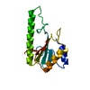
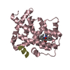
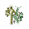
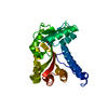
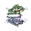
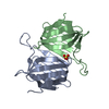
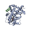
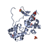
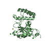
| Title | Very-short-patch DNA repair endonuclease bound to its reaction product site | ||||||
 Components Components |
| ||||||
 Keywords Keywords | HYDROLASE / DNA REPAIR / ENDONUCLEASE / VERY SHORT PATCH REPAIR / DNA REPAI HYDROLASE / NUCLEASE / ZINC / METAL-BINDING | ||||||
| Function / homology |  Function and homology information Function and homology informationT/G mismatch-specific endonuclease activity / mismatch repair / Hydrolases; Acting on ester bonds / hydrolase activity / DNA binding / metal ion binding Similarity search - Function | ||||||
| Biological species |  | ||||||
| Method |  X-RAY DIFFRACTION / SYNCHROTRON / MOLECULAR REPLACEMENT / Resolution: 2.8 Å X-RAY DIFFRACTION / SYNCHROTRON / MOLECULAR REPLACEMENT / Resolution: 2.8 Å | ||||||
 Authors Authors | Bunting, K.A. / Roe, S.M. / Headley, A. / Brown, T. / Savva, R. / Pearl, L.H. | ||||||
 Citation Citation | Journal: Nucleic Acids Res. / Year: 2003 Title: Crystal Structure of the Escherichia Coli Dcm Very-Short-Patch DNA Repair Endonuclease Bound to its Reaction Product-Site in a DNA Superhelix Authors: Bunting, K.A. / Roe, S.M. / Headley, A. / Brown, T. / Savva, R. / Pearl, L.H. | ||||||
| History |
|
- Structure visualization
| Structure viewer | Molecule: MolmilJmol/JSmol |
|---|
- Downloads & links
-Download
| PDBx/mmCIF format | 1odg.cif.gz | 59.4 KB | Display | PDBx/mmCIF format |
|---|---|---|---|---|
| PDB format | pdb1odg.ent.gz | 40.2 KB | Display | PDB format |
| PDBx/mmJSON format | 1odg.json.gz | Tree view | PDBx/mmJSON format | |
| Others |  Other downloads Other downloads |
-Validation report
| Arichive directory | https://data.pdbj.org/pub/pdb/validation_reports/od/1odgftp://data.pdbj.org/pub/pdb/validation_reports/od/1odg | HTTPS FTP |
|---|
-Related structure data
| Related structure data | 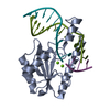 1cw0S S: Starting model for refinement |
|---|---|
| Similar structure data |


-Links
 PDBj
PDBj


- Assembly
| Deposited unit | 
| ||||||||
|---|---|---|---|---|---|---|---|---|---|
| 1 |
| ||||||||
| Unit cell |
| ||||||||
| Details | THE TRIMERIC ASSEMBLY DESCRIBED HERE IS FORMED BYTHE COMPLEX OF PROTEIN CHAIN A WITH DNA CHAINS F,W |
-Components
| #1: Protein | Mass: 15568.826 Da / Num. of mol.: 1 Source method: isolated from a genetically manipulated source Source: (gene. exp.) | ||||||
|---|---|---|---|---|---|---|---|
| #2: DNA chain | Mass: 3692.430 Da / Num. of mol.: 2 / Source method: isolated from a natural source / Source: (natural) #3: Chemical | ChemComp-ZN / |   Mass: 65.409 Da / Num. of mol.: 1 / Source method: obtained synthetically / Formula: Zn Mass: 65.409 Da / Num. of mol.: 1 / Source method: obtained synthetically / Formula: Zn#4: Water | ChemComp-HOH / |  Mass: 18.015 Da / Num. of mol.: 81 / Source method: isolated from a natural source / Formula: H2O Mass: 18.015 Da / Num. of mol.: 81 / Source method: isolated from a natural source / Formula: H2OCompound details | FUNCTIONS IN THE DEAMINATION OF 5-METHYLCYTOSINE IN DNA CAUSING MISMATCHES IN T/G WHICH CAN LEAD TO ...FUNCTIONS IN THE DEAMINATIO | |
-Experimental details
-Experiment
| Experiment | Method: X-RAY DIFFRACTION / Number of used crystals: 1 |
|---|
- Sample preparation
| Crystal | Density Matthews: 4.2 Å3/Da / Density % sol: 70.7 % | ||||||||||||||||||||||||||||||||||||||||||||||||||||||||
|---|---|---|---|---|---|---|---|---|---|---|---|---|---|---|---|---|---|---|---|---|---|---|---|---|---|---|---|---|---|---|---|---|---|---|---|---|---|---|---|---|---|---|---|---|---|---|---|---|---|---|---|---|---|---|---|---|---|
| Crystal grow | pH: 6.5 Details: 25 MM HEPES PH7.0, 75 MM NACL, 15% PEG 8000, 50 MM SODIUM CACODYLATE PH 6.5, 75 MM AMMONIUM SULPHATE AND 10% GLYCEROL PROTEIN 2.5 MG/ML | ||||||||||||||||||||||||||||||||||||||||||||||||||||||||
| Crystal grow | *PLUS Temperature: 14 ℃ / Method: batch method | ||||||||||||||||||||||||||||||||||||||||||||||||||||||||
| Components of the solutions | *PLUS
|
-Data collection
| Diffraction | Mean temperature: 100 K |
|---|---|
| Diffraction source | Source: SYNCHROTRON / Site: ESRF  / Beamline: ID14-2 / Wavelength: 0.933 / Beamline: ID14-2 / Wavelength: 0.933 |
| Detector | Type: ADSC CCD / Detector: CCD / Date: Feb 15, 2000 |
| Radiation | Protocol: SINGLE WAVELENGTH / Monochromatic (M) / Laue (L): M / Scattering type: x-ray |
| Radiation wavelength | Wavelength: 0.933 Å / Relative weight: 1 |
| Reflection | Resolution: 2.75→50 Å / Num. obs: 10084 / % possible obs: 99 % / Redundancy: 4.9 % / Biso Wilson estimate: 105 Å2 / Rmerge(I) obs: 0.07 / Net I/σ(I): 6.9 |
| Reflection shell | Resolution: 2.75→2.9 Å / Redundancy: 5 % / Rmerge(I) obs: 0.384 / Mean I/σ(I) obs: 2 / % possible all: 100 |
| Reflection | *PLUS Rmerge(I) obs: 0.07 |
| Reflection shell | *PLUS % possible obs: 100 % / Num. unique obs: 1464 |
- Processing
| Software |
| ||||||||||||||||||||||||||||||||||||||||||||||||||||||||||||||||||||||||||||||||
|---|---|---|---|---|---|---|---|---|---|---|---|---|---|---|---|---|---|---|---|---|---|---|---|---|---|---|---|---|---|---|---|---|---|---|---|---|---|---|---|---|---|---|---|---|---|---|---|---|---|---|---|---|---|---|---|---|---|---|---|---|---|---|---|---|---|---|---|---|---|---|---|---|---|---|---|---|---|---|---|---|---|
| Refinement | Method to determine structure: MOLECULAR REPLACEMENT Starting model: PDB ENTRY 1CW0 Resolution: 2.8→24.7 Å / Isotropic thermal model: RESTRAINED / Cross valid method: THROUGHOUT / σ(F): 0
| ||||||||||||||||||||||||||||||||||||||||||||||||||||||||||||||||||||||||||||||||
| Solvent computation | Solvent model: FLAT MODEL / Bsol: 74.25 Å2 / ksol: 0.318 e/Å3 | ||||||||||||||||||||||||||||||||||||||||||||||||||||||||||||||||||||||||||||||||
| Displacement parameters |
| ||||||||||||||||||||||||||||||||||||||||||||||||||||||||||||||||||||||||||||||||
| Refinement step | Cycle: LAST / Resolution: 2.8→24.7 Å
| ||||||||||||||||||||||||||||||||||||||||||||||||||||||||||||||||||||||||||||||||
| Refine LS restraints |
| ||||||||||||||||||||||||||||||||||||||||||||||||||||||||||||||||||||||||||||||||
| Xplor file |
| ||||||||||||||||||||||||||||||||||||||||||||||||||||||||||||||||||||||||||||||||
| Refinement | *PLUS Rfactor Rfree: 0.356 / Rfactor Rwork: 0.287 | ||||||||||||||||||||||||||||||||||||||||||||||||||||||||||||||||||||||||||||||||
| Solvent computation | *PLUS | ||||||||||||||||||||||||||||||||||||||||||||||||||||||||||||||||||||||||||||||||
| Displacement parameters | *PLUS | ||||||||||||||||||||||||||||||||||||||||||||||||||||||||||||||||||||||||||||||||
| Refine LS restraints | *PLUS
|
