 Movie
Movie Controller

[English] 日本語
 Yorodumi
- PDB-1obb: alpha-glucosidase A, AglA, from Thermotoga maritima in complex wi... -
+ Open data

- Basic information
| Entry | Database: PDB / ID: 1obb | |||||||||
|---|---|---|---|---|---|---|---|---|---|---|
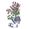
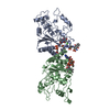
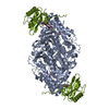
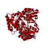
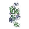
| Title | alpha-glucosidase A, AglA, from Thermotoga maritima in complex with maltose and NAD+ | |||||||||
 Components Components | ALPHA-GLUCOSIDASE | |||||||||
 Keywords Keywords | HYDROLASE / GLYCOSIDASE / SULFINIC ACID / NAD+ / MALTOSE | |||||||||
| Function / homology |  Function and homology information Function and homology informationalpha-glucosidase / alpha-1,4-glucosidase activity / oxidoreductase activity, acting on the CH-OH group of donors, NAD or NADP as acceptor / hydrolase activity, hydrolyzing O-glycosyl compounds / carbohydrate metabolic process / metal ion binding / cytosol Similarity search - Function | |||||||||
| Biological species |   THERMOTOGA MARITIMA (bacteria) THERMOTOGA MARITIMA (bacteria) | |||||||||
| Method |  X-RAY DIFFRACTION / MIRAS / Resolution: 1.9 Å X-RAY DIFFRACTION / MIRAS / Resolution: 1.9 Å | |||||||||
 Authors Authors | Lodge, J.A. / Maier, T. / Liebl, W. / Hoffmann, V. / Strater, N. | |||||||||
 Citation Citation | Journal: J.Biol.Chem. / Year: 2003 Title: Crystal Structure of Thermotoga Maritima Alpha-Glucosidase Agla Defines a New Clan of Nad+-Dependent Glycosidases Authors: Lodge, J.A. / Maier, T. / Liebl, W. / Hoffmann, V. / Strater, N. | |||||||||
| History |
| |||||||||
| Remark 700 | SHEET THE SHEET STRUCTURE OF THIS MOLECULE IS BIFURCATED. IN ORDER TO REPRESENT THIS FEATURE IN ... SHEET THE SHEET STRUCTURE OF THIS MOLECULE IS BIFURCATED. IN ORDER TO REPRESENT THIS FEATURE IN THE SHEET RECORDS BELOW, TWO SHEETS ARE DEFINED. |
- Structure visualization
| Structure viewer | Molecule: MolmilJmol/JSmol |
|---|
- Downloads & links
-Download
| PDBx/mmCIF format | 1obb.cif.gz | 217.3 KB | Display | PDBx/mmCIF format |
|---|---|---|---|---|
| PDB format | pdb1obb.ent.gz | 174.3 KB | Display | PDB format |
| PDBx/mmJSON format | 1obb.json.gz | Tree view | PDBx/mmJSON format | |
| Others |  Other downloads Other downloads |
-Validation report
| Arichive directory | https://data.pdbj.org/pub/pdb/validation_reports/ob/1obbftp://data.pdbj.org/pub/pdb/validation_reports/ob/1obb | HTTPS FTP |
|---|
-Related structure data
| Similar structure data |
|---|
-Links
 PDBj
PDBj


- Assembly
| Deposited unit | 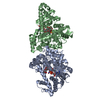
| ||||||||
|---|---|---|---|---|---|---|---|---|---|
| 1 |
| ||||||||
| Unit cell |
| ||||||||
| Noncrystallographic symmetry (NCS) | NCS oper: (Code: given Matrix: (-0.98427, 0.17666, 0.00361), Vector: |
-Components
| #1: Protein | Mass: 55154.078 Da / Num. of mol.: 2 Source method: isolated from a genetically manipulated source Source: (gene. exp.) THERMOTOGA MARITIMA (bacteria) / Strain: MSB8 / Plasmid: PUN121 / Production host: #2: Polysaccharide |   Source method: isolated from a genetically manipulated source Details: oligosaccharide / References: alpha-maltose #3: Chemical |   Mass: 663.425 Da / Num. of mol.: 2 / Source method: obtained synthetically / Formula: C21H27N7O14P2 / Comment: NAD*YM Mass: 663.425 Da / Num. of mol.: 2 / Source method: obtained synthetically / Formula: C21H27N7O14P2 / Comment: NAD*YM#4: Water | ChemComp-HOH / |  Mass: 18.015 Da / Num. of mol.: 646 / Source method: isolated from a natural source / Formula: H2O Mass: 18.015 Da / Num. of mol.: 646 / Source method: isolated from a natural source / Formula: H2OHas protein modification | Y | |
|---|
-Experimental details
-Experiment
| Experiment | Method: X-RAY DIFFRACTION / Number of used crystals: 1 |
|---|
- Sample preparation
| Crystal | Density Matthews: 2.586 Å3/Da / Density % sol: 50.6 % | |||||||||||||||||||||||||||||||||||||||||||||||||
|---|---|---|---|---|---|---|---|---|---|---|---|---|---|---|---|---|---|---|---|---|---|---|---|---|---|---|---|---|---|---|---|---|---|---|---|---|---|---|---|---|---|---|---|---|---|---|---|---|---|---|
| Crystal grow | pH: 5 Details: 10% PEG 6000, 1M LICL, 0.1M TRIS/HCL PH4.6, 2MM MNCL2, 50MM MALTOSE, 50MM NAD+, pH 5.00 | |||||||||||||||||||||||||||||||||||||||||||||||||
| Crystal grow | *PLUS pH: 7 / Method: vapor diffusion, hanging drop | |||||||||||||||||||||||||||||||||||||||||||||||||
| Components of the solutions | *PLUS
|
-Data collection
| Diffraction | Mean temperature: 100 K |
|---|---|
| Diffraction source | Source: ROTATING ANODE / Type: ENRAF-NONIUS FR571 / Wavelength: 1.5475 |
| Detector | Type: MAR scanner 345 mm plate / Detector: IMAGE PLATE / Date: Nov 15, 2001 / Details: OSMIC MULTILAYER MAX-FLUX |
| Radiation | Monochromator: OSMIC MULTILAYER MAX-FLUX / Protocol: SINGLE WAVELENGTH / Monochromatic (M) / Laue (L): M / Scattering type: x-ray |
| Radiation wavelength | Wavelength: 1.5475 Å / Relative weight: 1 |
| Reflection | Resolution: 1.9→24 Å / Num. obs: 77496 / % possible obs: 98.3 % / Redundancy: 2.6 % / Biso Wilson estimate: 21.4 Å2 / Rmerge(I) obs: 0.045 / Net I/σ(I): 16.9 |
| Reflection shell | Resolution: 1.9→1.99 Å / Redundancy: 2.5 % / Rmerge(I) obs: 0.396 / Mean I/σ(I) obs: 2.4 / % possible all: 93.7 |
| Reflection | *PLUS Highest resolution: 1.9 Å / Lowest resolution: 25 Å / % possible obs: 96.6 % / Redundancy: 2.5 % / Rmerge(I) obs: 0.038 |
| Reflection shell | *PLUS Lowest resolution: 1.97 Å / % possible obs: 92.6 % / Redundancy: 2.4 % / Rmerge(I) obs: 0.362 / Mean I/σ(I) obs: 2.1 |
- Processing
| Software |
| ||||||||||||||||||||||||||||||||||||||||||||||||||||||||||||||||||||||||||||||||
|---|---|---|---|---|---|---|---|---|---|---|---|---|---|---|---|---|---|---|---|---|---|---|---|---|---|---|---|---|---|---|---|---|---|---|---|---|---|---|---|---|---|---|---|---|---|---|---|---|---|---|---|---|---|---|---|---|---|---|---|---|---|---|---|---|---|---|---|---|---|---|---|---|---|---|---|---|---|---|---|---|---|
| Refinement | Method to determine structure: MIRAS / Resolution: 1.9→17.65 Å / Rfactor Rfree error: 0.005 / Data cutoff high absF: 1732125.74 / Data cutoff low absF: 0 / Isotropic thermal model: RESTRAINED / Cross valid method: THROUGHOUT / σ(F): 0
| ||||||||||||||||||||||||||||||||||||||||||||||||||||||||||||||||||||||||||||||||
| Solvent computation | Solvent model: FLAT MODEL / Bsol: 57.5501 Å2 / ksol: 0.378793 e/Å3 | ||||||||||||||||||||||||||||||||||||||||||||||||||||||||||||||||||||||||||||||||
| Displacement parameters | Biso mean: 32.5 Å2
| ||||||||||||||||||||||||||||||||||||||||||||||||||||||||||||||||||||||||||||||||
| Refine analyze |
| ||||||||||||||||||||||||||||||||||||||||||||||||||||||||||||||||||||||||||||||||
| Refinement step | Cycle: LAST / Resolution: 1.9→17.65 Å
| ||||||||||||||||||||||||||||||||||||||||||||||||||||||||||||||||||||||||||||||||
| Refine LS restraints |
| ||||||||||||||||||||||||||||||||||||||||||||||||||||||||||||||||||||||||||||||||
| LS refinement shell | Resolution: 1.9→2.02 Å / Rfactor Rfree error: 0.016 / Total num. of bins used: 6
| ||||||||||||||||||||||||||||||||||||||||||||||||||||||||||||||||||||||||||||||||
| Xplor file |
| ||||||||||||||||||||||||||||||||||||||||||||||||||||||||||||||||||||||||||||||||
| Refinement | *PLUS Rfactor Rfree: 0.234 / Rfactor Rwork: 0.196 | ||||||||||||||||||||||||||||||||||||||||||||||||||||||||||||||||||||||||||||||||
| Solvent computation | *PLUS | ||||||||||||||||||||||||||||||||||||||||||||||||||||||||||||||||||||||||||||||||
| Displacement parameters | *PLUS | ||||||||||||||||||||||||||||||||||||||||||||||||||||||||||||||||||||||||||||||||
| Refine LS restraints | *PLUS
| ||||||||||||||||||||||||||||||||||||||||||||||||||||||||||||||||||||||||||||||||
| LS refinement shell | *PLUS Lowest resolution: 1.97 Å |
