 Movie
Movie Controller

[English] 日本語
 Yorodumi
- PDB-1o9y: Crystal structure of the C-terminal domain of the HrcQb protein f... -
+ Open data

- Basic information
| Entry | Database: PDB / ID: 1o9y | ||||||
|---|---|---|---|---|---|---|---|
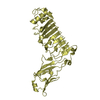
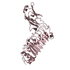
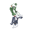
| Title | Crystal structure of the C-terminal domain of the HrcQb protein from Pseudomonas syringae pv. phaseolicola | ||||||
 Components Components | HRCQ2 | ||||||
 Keywords Keywords | STRUCTURAL PROTEIN / SECRETORY PROTEIN / HRP / TYPE III SECRETION SYSTEM / PHYTOPATHOGENICITY | ||||||
| Function / homology |  Function and homology information Function and homology informationbacterial-type flagellum basal body / cytoskeletal motor activity / bacterial-type flagellum-dependent cell motility / chemotaxis / cytoplasm Similarity search - Function | ||||||
| Biological species |  PSEUDOMONAS SYRINGAE (bacteria) PSEUDOMONAS SYRINGAE (bacteria) | ||||||
| Method |  X-RAY DIFFRACTION / SYNCHROTRON / MAD / Resolution: 2.29 Å X-RAY DIFFRACTION / SYNCHROTRON / MAD / Resolution: 2.29 Å | ||||||
 Authors Authors | Fadouloglou, V.E. / Kokkinidis, M. | ||||||
 Citation Citation | Journal: Proc.Natl.Acad.Sci.USA / Year: 2004 Title: Structure of Hrcqb-C, a Conserved Component of the Bacterial Type III Secretion Systems. Authors: Fadouloglou, V.E. / Tampakaki, A.P. / Glykos, N.M. / Bastaki, M.N. / Hadden, J.M. / Phillips, S.E. / Panopoulos, N.J. / Kokkinidis, M. #1: Journal: Acta Crystallogr.,Sect.D / Year: 2001 Title: Structural Studies of the Hrp Secretion System: Expression, Purification, Crystallization and Preliminary X-Ray Analysis of the C-Terminal Domain of the Hrcqb Protein from Pseudomonas Syringae Pv. Phaseolicola. Authors: Fadouloglou, V.E. / Tampakaki, A.P. / Panopoulos, N.J. / Kokkinidis, M. | ||||||
| History |
|
- Structure visualization
| Structure viewer | Molecule: MolmilJmol/JSmol |
|---|
- Downloads & links
-Download
| PDBx/mmCIF format | 1o9y.cif.gz | 64.8 KB | Display | PDBx/mmCIF format |
|---|---|---|---|---|
| PDB format | pdb1o9y.ent.gz | 49.5 KB | Display | PDB format |
| PDBx/mmJSON format | 1o9y.json.gz | Tree view | PDBx/mmJSON format | |
| Others |  Other downloads Other downloads |
-Validation report
| Arichive directory | https://data.pdbj.org/pub/pdb/validation_reports/o9/1o9yftp://data.pdbj.org/pub/pdb/validation_reports/o9/1o9y | HTTPS FTP |
|---|
-Related structure data
| Similar structure data |
|---|


-Links
 PDBj
PDBj- Assembly
| Deposited unit | 
| ||||||||||||||||
|---|---|---|---|---|---|---|---|---|---|---|---|---|---|---|---|---|---|
| 1 |
| ||||||||||||||||
| Unit cell |
| ||||||||||||||||
| Noncrystallographic symmetry (NCS) | NCS oper:
| ||||||||||||||||
| Details | EVIDENCE BOTH FROM GEL FILTRATION EXPERIMENTS AND THECRYSTAL STRUCTURE SUGGESTS THAT THE TETRAMER (DIMER OFDIMERS) CONSISTING OF ALL FOUR CHAINS MAY BE BIOLOGICALLYRELEVANT. |
-Components
| #1: Protein | Mass: 8953.182 Da / Num. of mol.: 4 / Fragment: RESIDUES 50-128 Source method: isolated from a genetically manipulated source Source: (gene. exp.) PSEUDOMONAS SYRINGAE (bacteria)Description: THE C-TERMINUS (RESIDUES GLN 50 - SER 128) OF THE WHOLE PROTEIN WAS CLONED Variant: PHASEOLICOLA / Production host: #2: Water | ChemComp-HOH / |  Mass: 18.015 Da / Num. of mol.: 153 / Source method: isolated from a natural source / Formula: H2O Mass: 18.015 Da / Num. of mol.: 153 / Source method: isolated from a natural source / Formula: H2OHas protein modification | Y | |
|---|
-Experimental details
-Experiment
| Experiment | Method: X-RAY DIFFRACTION / Number of used crystals: 1 |
|---|
- Sample preparation
| Crystal | Density Matthews: 2.34 Å3/Da / Density % sol: 46.96 % | ||||||||||||||||||||||||||||||||||||||||||
|---|---|---|---|---|---|---|---|---|---|---|---|---|---|---|---|---|---|---|---|---|---|---|---|---|---|---|---|---|---|---|---|---|---|---|---|---|---|---|---|---|---|---|---|
| Crystal grow | pH: 6.5 Details: 15% V/V MPD, 80 MM MAGNESIUM ACETATE, 100 MM BIS-TRIS PH=6.5, pH 6.50 | ||||||||||||||||||||||||||||||||||||||||||
| Crystal grow | *PLUS Temperature: 292 K / pH: 6.5 / Method: vapor diffusion, hanging drop / Details: Fadouloglou, V.E., (2001) Acta Cryst., D57, 1689. | ||||||||||||||||||||||||||||||||||||||||||
| Components of the solutions | *PLUS
|
-Data collection
| Diffraction | Mean temperature: 100 K | ||||||||||||
|---|---|---|---|---|---|---|---|---|---|---|---|---|---|
| Diffraction source | Source: SYNCHROTRON / Site: ESRF  / Beamline: BM14 / Wavelength: 0.9800,0.9804,0.8856 / Beamline: BM14 / Wavelength: 0.9800,0.9804,0.8856 | ||||||||||||
| Detector | Type: MARRESEARCH / Detector: CCD / Date: Mar 15, 2002 | ||||||||||||
| Radiation | Protocol: MAD / Monochromatic (M) / Laue (L): M / Scattering type: x-ray | ||||||||||||
| Radiation wavelength |
| ||||||||||||
| Reflection | Resolution: 2.29→39 Å / Num. obs: 13028 / % possible obs: 98.5 % / Redundancy: 3.5 % / Biso Wilson estimate: 29 Å2 / Rsym value: 0.058 / Net I/σ(I): 9.3 | ||||||||||||
| Reflection shell | Resolution: 2.29→2.42 Å / Redundancy: 1.8 % / Mean I/σ(I) obs: 4.6 / Rsym value: 0.156 / % possible all: 88.6 | ||||||||||||
| Reflection | *PLUS Highest resolution: 2.3 Å / Lowest resolution: 39 Å / Num. obs: 12352 / % possible obs: 0.058 % / Redundancy: 3.4 % | ||||||||||||
| Reflection shell | *PLUS Highest resolution: 2.3 Å / % possible obs: 88.6 % / Redundancy: 1.8 % / Rmerge(I) obs: 0.156 / Mean I/σ(I) obs: 4.6 |
- Processing
| Software |
| ||||||||||||||||||||
|---|---|---|---|---|---|---|---|---|---|---|---|---|---|---|---|---|---|---|---|---|---|
| Refinement | Method to determine structure: MAD / Resolution: 2.29→39 Å / SU B: 6.373 / SU ML: 0.157 / Cross valid method: THROUGHOUT / ESU R: 0.34 / ESU R Free: 0.227 / Details: CNS
| ||||||||||||||||||||
| Displacement parameters | Biso mean: 22.682 Å2
| ||||||||||||||||||||
| Refinement step | Cycle: LAST / Resolution: 2.29→39 Å
| ||||||||||||||||||||
| Refinement | *PLUS Highest resolution: 2.3 Å / Rfactor Rfree: 0.228 / Rfactor Rwork: 0.182 | ||||||||||||||||||||
| Solvent computation | *PLUS | ||||||||||||||||||||
| Displacement parameters | *PLUS | ||||||||||||||||||||
| Refine LS restraints | *PLUS
|
