 Movie
Movie Controller


+ Open data

- Basic information
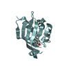
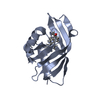
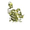
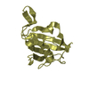
| Entry | Database: PDB / ID: 1o7i | ||||||
|---|---|---|---|---|---|---|---|
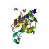
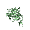
| Title | Crystal structure of a single stranded DNA binding protein | ||||||
 Components Components | SINGLE STRANDED DNA BINDING PROTEIN | ||||||
 Keywords Keywords | DNA BINDING PROTEIN / SINGLE STRANDED DNA / OB FOLD / DNA-BINDING PROTEIN | ||||||
| Function / homology |  Function and homology information Function and homology informationresponse to ionizing radiation / double-strand break repair via homologous recombination / chromosome / DNA binding / cytoplasm Similarity search - Function | ||||||
| Biological species |   SULFOLOBUS SOLFATARICUS (archaea) SULFOLOBUS SOLFATARICUS (archaea) | ||||||
| Method |  X-RAY DIFFRACTION / SYNCHROTRON / MAD / Resolution: 1.2 Å X-RAY DIFFRACTION / SYNCHROTRON / MAD / Resolution: 1.2 Å | ||||||
 Authors Authors | Kerr, I.D. / Naismith, J.H. | ||||||
 Citation Citation | Journal: EMBO J. / Year: 2003 Title: Insights into ssDNA recognition by the OB fold from a structural and thermodynamic study of Sulfolobus SSB protein. Authors: Kerr, I.D. / Wadsworth, R.I. / Cubeddu, L. / Blankenfeldt, W. / Naismith, J.H. / White, M.F. #1: Journal: Acta Crystallogr.,Sect.D / Year: 2001 Title: Overexpression, Purification, Crystallization and Data Collection of a Single-Stranded DNA-Binding Protein from Sulfolobus Solfataricus Authors: Kerr, I.D. / Wadsworth, R.I. / Blankenfeldt, W. / Staines, A.G. / White, M.F. / Naismith, J.H. | ||||||
| History |
| ||||||
| Remark 700 | SHEET THE SHEET STRUCTURE OF THIS MOLECULE IS BIFURCATED. IN ORDER TO REPRESENT THIS FEATURE IN ... SHEET THE SHEET STRUCTURE OF THIS MOLECULE IS BIFURCATED. IN ORDER TO REPRESENT THIS FEATURE IN THE SHEET RECORDS BELOW, TWO SHEETS ARE DEFINED. |
- Structure visualization
| Structure viewer | Molecule: MolmilJmol/JSmol |
|---|
- Downloads & links
-Download
| PDBx/mmCIF format | 1o7i.cif.gz | 107.2 KB | Display | PDBx/mmCIF format |
|---|---|---|---|---|
| PDB format | pdb1o7i.ent.gz | 83.5 KB | Display | PDB format |
| PDBx/mmJSON format | 1o7i.json.gz | Tree view | PDBx/mmJSON format | |
| Others |  Other downloads Other downloads |
-Validation report
| Arichive directory | https://data.pdbj.org/pub/pdb/validation_reports/o7/1o7iftp://data.pdbj.org/pub/pdb/validation_reports/o7/1o7i | HTTPS FTP |
|---|
-Related structure data
| Similar structure data |
|---|


-Links
 PDBj
PDBj- Assembly
| Deposited unit | 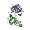
| ||||||||
|---|---|---|---|---|---|---|---|---|---|
| 1 | 
| ||||||||
| 2 | 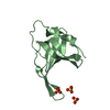
| ||||||||
| Unit cell |
| ||||||||
| Noncrystallographic symmetry (NCS) | NCS oper: (Code: given Matrix: (-0.997569, 0.042527, 0.05521), Vector: |
-Components
| #1: Protein | Mass: 12949.548 Da / Num. of mol.: 2 / Fragment: RESIDUES 1-119 Source method: isolated from a genetically manipulated source Source: (gene. exp.) SULFOLOBUS SOLFATARICUS (archaea) / Strain: P2 / Plasmid: PET19B / Production host:  #2: Chemical |   Mass: 96.063 Da / Num. of mol.: 3 / Source method: obtained synthetically / Formula: SO4 Mass: 96.063 Da / Num. of mol.: 3 / Source method: obtained synthetically / Formula: SO4#3: Water | ChemComp-HOH / |  Mass: 18.015 Da / Num. of mol.: 271 / Source method: isolated from a natural source / Formula: H2O Mass: 18.015 Da / Num. of mol.: 271 / Source method: isolated from a natural source / Formula: H2OCompound details | CONTAINS OB-FOLD DOMAINS THAT BIND TO NUCLEIC ACIDS. | |
|---|
-Experimental details
-Experiment
| Experiment | Method: X-RAY DIFFRACTION / Number of used crystals: 1 |
|---|
- Sample preparation
| Crystal | Density Matthews: 2.25 Å3/Da / Density % sol: 45 % | ||||||||||||||||||||||||
|---|---|---|---|---|---|---|---|---|---|---|---|---|---|---|---|---|---|---|---|---|---|---|---|---|---|
| Crystal grow | Temperature: 298 K / Method: vapor diffusion, hanging drop / pH: 5 Details: CITRIC ACID, AMMONIUM SULFATE, PH 5.0, VAPOUR DIFFUSION, HANGING DROP AT 298K | ||||||||||||||||||||||||
| Crystal grow | *PLUS Temperature: 298 K / Method: vapor diffusion, hanging dropDetails: Kerr, I.D., (2001) Acta Crystallogr.,Sect.D, 57, 1290. PH range low: 5 / PH range high: 3.5 | ||||||||||||||||||||||||
| Components of the solutions | *PLUS
|
-Data collection
| Diffraction | Mean temperature: 130 K | ||||||||||||
|---|---|---|---|---|---|---|---|---|---|---|---|---|---|
| Diffraction source | Source: SYNCHROTRON / Site: SRS  / Beamline: PX14.2 / Wavelength: 0.9600, 0.9780, 0.9786 / Beamline: PX14.2 / Wavelength: 0.9600, 0.9780, 0.9786 | ||||||||||||
| Detector | Type: ADSC CCD / Detector: CCD / Date: Mar 15, 2002 / Details: MIRRORS | ||||||||||||
| Radiation | Monochromator: SI INTERCHANGEABLE / Protocol: MAD / Monochromatic (M) / Laue (L): M / Scattering type: x-ray | ||||||||||||
| Radiation wavelength |
| ||||||||||||
| Reflection | Resolution: 1.69→37.3 Å / Num. obs: 24609 / % possible obs: 100 % / Redundancy: 16.6 % / Rmerge(I) obs: 0.054 / Net I/σ(I): 8.1 | ||||||||||||
| Reflection shell | Resolution: 1.69→1.73 Å / Redundancy: 16.5 % / Rmerge(I) obs: 0.352 / Mean I/σ(I) obs: 2.2 / % possible all: 100 | ||||||||||||
| Reflection | *PLUS Highest resolution: 1.26 Å / Lowest resolution: 47.92 Å / Num. obs: 60733 / % possible obs: 99.9 % / Redundancy: 7.5 % / Num. measured all: 463924 / Rmerge(I) obs: 0.081 | ||||||||||||
| Reflection shell | *PLUS Highest resolution: 1.26 Å / Lowest resolution: 1.29 Å / % possible obs: 99.9 % / Redundancy: 3.6 % / Rmerge(I) obs: 0.352 / Mean I/σ(I) obs: 2 |
- Processing
| Software |
| ||||||||||||||||||||||||||||||||||||||||||||||||||||||||||||||||||||||||||||||||||||||||||||||||||||||||||||||||||||||||||||||||||||||||||||||||||||||||||||||||||||||||||||||||||||||
|---|---|---|---|---|---|---|---|---|---|---|---|---|---|---|---|---|---|---|---|---|---|---|---|---|---|---|---|---|---|---|---|---|---|---|---|---|---|---|---|---|---|---|---|---|---|---|---|---|---|---|---|---|---|---|---|---|---|---|---|---|---|---|---|---|---|---|---|---|---|---|---|---|---|---|---|---|---|---|---|---|---|---|---|---|---|---|---|---|---|---|---|---|---|---|---|---|---|---|---|---|---|---|---|---|---|---|---|---|---|---|---|---|---|---|---|---|---|---|---|---|---|---|---|---|---|---|---|---|---|---|---|---|---|---|---|---|---|---|---|---|---|---|---|---|---|---|---|---|---|---|---|---|---|---|---|---|---|---|---|---|---|---|---|---|---|---|---|---|---|---|---|---|---|---|---|---|---|---|---|---|---|---|---|
| Refinement | Method to determine structure: MAD / Resolution: 1.2→65 Å / Cor.coef. Fo:Fc: 0.966 / Cor.coef. Fo:Fc free: 0.954 / SU B: 0.805 / SU ML: 0.035 / Cross valid method: THROUGHOUT / ESU R: 0.047 / ESU R Free: 0.046 / Stereochemistry target values: MAXIMUM LIKELIHOOD Details: HYDROGENS HAVE BEEN ADDED IN THE RIDING POSITIONS.RESIDUES 116-119 OF CHAIN A & 115-119 OF CHAIN B WERE NOT LOCATED IN THE ELECTRON DENSITY AND THEREFORE NOT INCLUDED IN THE MODEL. ATOMS AT ...Details: HYDROGENS HAVE BEEN ADDED IN THE RIDING POSITIONS.RESIDUES 116-119 OF CHAIN A & 115-119 OF CHAIN B WERE NOT LOCATED IN THE ELECTRON DENSITY AND THEREFORE NOT INCLUDED IN THE MODEL. ATOMS AT 0.01 OCCUPANCY WERE NOT LOCATED ON THE ELECTRON DENSITY AND MODELLED STEREOCHEMICALLY.
| ||||||||||||||||||||||||||||||||||||||||||||||||||||||||||||||||||||||||||||||||||||||||||||||||||||||||||||||||||||||||||||||||||||||||||||||||||||||||||||||||||||||||||||||||||||||
| Solvent computation | Ion probe radii: 0.8 Å / Shrinkage radii: 0.8 Å / VDW probe radii: 1.4 Å / Solvent model: BABINET MODEL WITH MASK | ||||||||||||||||||||||||||||||||||||||||||||||||||||||||||||||||||||||||||||||||||||||||||||||||||||||||||||||||||||||||||||||||||||||||||||||||||||||||||||||||||||||||||||||||||||||
| Displacement parameters | Biso mean: 10.73 Å2
| ||||||||||||||||||||||||||||||||||||||||||||||||||||||||||||||||||||||||||||||||||||||||||||||||||||||||||||||||||||||||||||||||||||||||||||||||||||||||||||||||||||||||||||||||||||||
| Refinement step | Cycle: LAST / Resolution: 1.2→65 Å
| ||||||||||||||||||||||||||||||||||||||||||||||||||||||||||||||||||||||||||||||||||||||||||||||||||||||||||||||||||||||||||||||||||||||||||||||||||||||||||||||||||||||||||||||||||||||
| Refine LS restraints |
|