 Movie
Movie Controller


+ Open data

- Basic information
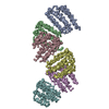
| Entry | Database: PDB / ID: 1o6e | ||||||
|---|---|---|---|---|---|---|---|
| Title | Epstein-Barr virus protease | ||||||
 Components Components | CAPSID PROTEIN P40 | ||||||
 Keywords Keywords | HYDROLASE / PROTEINASE / BETA-BARREL / SERINE PROTEASE / STRUCTURAL PROTEOMICS IN EUROPE / SPINE / STRUCTURAL GENOMICS | ||||||
| Function / homology |  Function and homology information Function and homology informationassemblin / nuclear capsid assembly / viral release from host cell / host cell cytoplasm / serine-type endopeptidase activity / host cell nucleus / proteolysis / identical protein binding Similarity search - Function | ||||||
| Biological species |  HUMAN HERPESVIRUS 4 (Epstein-Barr virus) HUMAN HERPESVIRUS 4 (Epstein-Barr virus) | ||||||
| Method |  X-RAY DIFFRACTION / SYNCHROTRON / MOLECULAR REPLACEMENT / Resolution: 2.3 Å X-RAY DIFFRACTION / SYNCHROTRON / MOLECULAR REPLACEMENT / Resolution: 2.3 Å | ||||||
 Authors Authors | Buisson, M. / Hernandez, J. / Lascoux, D. / Schoehn, G. / Forest, E. / Arlaud, G. / Seigneurin, J. / Ruigrok, R.W.H. / Burmeister, W.P. | ||||||
 Citation Citation | Journal: J.Mol.Biol. / Year: 2002 Title: The Crystal Structure of the Epstein-Barr Virus Protease Shows Rearrangement of the Processed C Terminus Authors: Buisson, M. / Hernandez, J. / Lascoux, D. / Schoehn, G. / Forest, E. / Arlaud, G. / Seigneurin, J. / Ruigrok, R.W.H. / Burmeister, W.P. | ||||||
| History |
|
- Structure visualization
| Structure viewer | Molecule: MolmilJmol/JSmol |
|---|
- Downloads & links
-Download
| PDBx/mmCIF format | 1o6e.cif.gz | 99.1 KB | Display | PDBx/mmCIF format |
|---|---|---|---|---|
| PDB format | pdb1o6e.ent.gz | 76.8 KB | Display | PDB format |
| PDBx/mmJSON format | 1o6e.json.gz | Tree view | PDBx/mmJSON format | |
| Others |  Other downloads Other downloads |
-Validation report
| Arichive directory | https://data.pdbj.org/pub/pdb/validation_reports/o6/1o6eftp://data.pdbj.org/pub/pdb/validation_reports/o6/1o6e | HTTPS FTP |
|---|
-Related structure data
| Related structure data |  1fl1S S: Starting model for refinement |
|---|---|
| Similar structure data |


-Links
 PDBj
PDBj- Assembly
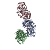
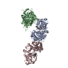
| Deposited unit | 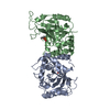
| ||||||||
|---|---|---|---|---|---|---|---|---|---|
| 1 | 
| ||||||||
| Unit cell |
| ||||||||
| Noncrystallographic symmetry (NCS) | NCS oper: (Code: given Matrix: (-0.9993, 0.0239, 0.0285), Vector: |
-Components
| #1: Protein | Mass: 25423.990 Da / Num. of mol.: 2 Fragment: COAT PROTEIN VP24 (PROTEASE) DOMAIN, RESIDUES 1-235 Mutation: YES Source method: isolated from a genetically manipulated source Source: (gene. exp.) HUMAN HERPESVIRUS 4 (Epstein-Barr virus)Strain: B95-8 / Cell line: B95-8 MARMOSET CELL LINE / Plasmid: PET28A / Production host:  #2: Chemical |   Mass: 140.075 Da / Num. of mol.: 2 / Source method: obtained synthetically / Formula: C3H9O4P Mass: 140.075 Da / Num. of mol.: 2 / Source method: obtained synthetically / Formula: C3H9O4P#3: Water | ChemComp-HOH / |  Mass: 18.015 Da / Num. of mol.: 12 / Source method: isolated from a natural source / Formula: H2O Mass: 18.015 Da / Num. of mol.: 12 / Source method: isolated from a natural source / Formula: H2OCompound details | ENGINEERED | Has protein modification | Y | Sequence details | ONLY PROTEASE DOMAIN OF THE ASSEMBLIN IS PRESENT IN THE CRYSTAL.THE SEQUENCE IS MODIFIED AT THE N- ...ONLY PROTEASE DOMAIN OF THE ASSEMBLIN IS PRESENT IN THE CRYSTAL.THE SEQUENCE IS MODIFIED AT THE N-TERMINUS DUE TO THE INTRODUCTI | |
|---|
-Experimental details
-Experiment
| Experiment | Method: X-RAY DIFFRACTION / Number of used crystals: 1 |
|---|
- Sample preparation
| Crystal | Density Matthews: 2.57 Å3/Da / Density % sol: 52 % / Description: CRYSTAL TWINNED | ||||||||||||||||||||||||||||||||||||||||||||||||||||||||||||||||||||||
|---|---|---|---|---|---|---|---|---|---|---|---|---|---|---|---|---|---|---|---|---|---|---|---|---|---|---|---|---|---|---|---|---|---|---|---|---|---|---|---|---|---|---|---|---|---|---|---|---|---|---|---|---|---|---|---|---|---|---|---|---|---|---|---|---|---|---|---|---|---|---|---|
| Crystal grow | Method: vapor diffusion, hanging drop / pH: 4.6 Details: HANGING DROP METHOD USING A RESERVOIR SOLUTION OF 1.25 - 1.5 M SODIUM FORMATE,100 MM SODIUM CITRATE PH4.6 5 MM EDTA, PROTEIN IN 100 MM NACL 20 MM THRIS-HCL PH 7.5 1 MM EDTA 10 MM BETA-MERCAPTOETHANOL | ||||||||||||||||||||||||||||||||||||||||||||||||||||||||||||||||||||||
| Crystal grow | *PLUS pH: 7.5 / Method: vapor diffusion, hanging drop | ||||||||||||||||||||||||||||||||||||||||||||||||||||||||||||||||||||||
| Components of the solutions | *PLUS
|
-Data collection
| Diffraction | Mean temperature: 100 K |
|---|---|
| Diffraction source | Source: SYNCHROTRON / Site: ESRF  / Beamline: ID14-4 / Wavelength: 0.919 / Beamline: ID14-4 / Wavelength: 0.919 |
| Detector | Type: ADSC CCD / Detector: CCD / Date: Jun 15, 2001 / Details: TOROIDAL MIRROR |
| Radiation | Monochromator: SI(111) / Protocol: SINGLE WAVELENGTH / Monochromatic (M) / Laue (L): M / Scattering type: x-ray |
| Radiation wavelength | Wavelength: 0.919 Å / Relative weight: 1 |
| Reflection | Resolution: 2.3→45.2 Å / Num. obs: 132854 / % possible obs: 98.1 % / Redundancy: 5.4 % / Biso Wilson estimate: 56.8 Å2 / Rmerge(I) obs: 0.081 / Net I/σ(I): 5.2 |
| Reflection shell | Resolution: 2.3→2.38 Å / Redundancy: 3.2 % / Rmerge(I) obs: 0.319 / Mean I/σ(I) obs: 2.3 / % possible all: 89.9 |
| Reflection shell | *PLUS Highest resolution: 2.3 Å / % possible obs: 89.9 % |
- Processing
| Software |
| ||||||||||||||||||||||||||||||||||||||||||||||||||||||||||||||||||||||||||||||||
|---|---|---|---|---|---|---|---|---|---|---|---|---|---|---|---|---|---|---|---|---|---|---|---|---|---|---|---|---|---|---|---|---|---|---|---|---|---|---|---|---|---|---|---|---|---|---|---|---|---|---|---|---|---|---|---|---|---|---|---|---|---|---|---|---|---|---|---|---|---|---|---|---|---|---|---|---|---|---|---|---|---|
| Refinement | Method to determine structure: MOLECULAR REPLACEMENT Starting model: PDB ENTRY 1FL1 Resolution: 2.3→42 Å / Rfactor Rfree error: 0.008 / Data cutoff high absF: 0 / Isotropic thermal model: RESTRAINED / Cross valid method: THROUGHOUT / σ(F): 0 Details: REFINED USING THE TWINNED TARGET, TWINNING FRACTION 0.477, OPERATOR -H,-K,L THE REFINEMENT HAS BEEN CARRIED OUT AGAINST TWINNED DATA USING THE CORRESPONDING TARGET IN CNS. R-FACTORS CAN NOT ...Details: REFINED USING THE TWINNED TARGET, TWINNING FRACTION 0.477, OPERATOR -H,-K,L THE REFINEMENT HAS BEEN CARRIED OUT AGAINST TWINNED DATA USING THE CORRESPONDING TARGET IN CNS. R-FACTORS CAN NOT DIRECTLY BE COMPARED TO THE ONES FROM UNTWINNED STRUCTURES. FOR MAP CALCULATIONS, A DIFFERENT DATASET WITH A TWINNING FRACTION MORE DIFFERENT FROM 0.5 HAS BEEN USED. ITS RESOLUTION WAS LIMITED TO 2.5 A. SOME REGIONS CARACTERISED BY B FACTORS ABOVE 90 A2 ARE POORLY DEFINED.
| ||||||||||||||||||||||||||||||||||||||||||||||||||||||||||||||||||||||||||||||||
| Solvent computation | Solvent model: FLAT MODEL / Bsol: 26.6 Å2 / ksol: 0.313 e/Å3 | ||||||||||||||||||||||||||||||||||||||||||||||||||||||||||||||||||||||||||||||||
| Displacement parameters | Biso mean: 66.1 Å2
| ||||||||||||||||||||||||||||||||||||||||||||||||||||||||||||||||||||||||||||||||
| Refine analyze |
| ||||||||||||||||||||||||||||||||||||||||||||||||||||||||||||||||||||||||||||||||
| Refinement step | Cycle: LAST / Resolution: 2.3→42 Å
| ||||||||||||||||||||||||||||||||||||||||||||||||||||||||||||||||||||||||||||||||
| Refine LS restraints |
| ||||||||||||||||||||||||||||||||||||||||||||||||||||||||||||||||||||||||||||||||
| LS refinement shell | Resolution: 2.3→2.38 Å / Rfactor Rfree error: 0.038 / Total num. of bins used: 10
| ||||||||||||||||||||||||||||||||||||||||||||||||||||||||||||||||||||||||||||||||
| Xplor file |
| ||||||||||||||||||||||||||||||||||||||||||||||||||||||||||||||||||||||||||||||||
| Refine LS restraints | *PLUS
|
