 Movie
Movie Controller

[English] 日本語
 Yorodumi
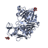
- PDB-1o5l: Crystal structure of Transcriptional regulator (TM1171) from Ther... -
+ Open data

- Basic information
| Entry | Database: PDB / ID: 1o5l | ||||||
|---|---|---|---|---|---|---|---|
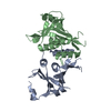
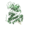
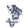
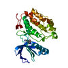
| Title | Crystal structure of Transcriptional regulator (TM1171) from Thermotoga maritima at 2.30 A resolution | ||||||
 Components Components | transcriptional regulator, crp family | ||||||
 Keywords Keywords | TRANSCRIPTION / TM1171 / TRANSCRIPTIONAL REGULATOR / CRP FAMILY / STRUCTURAL GENOMICS / JCSG / PSI / Protein Structure Initiative / Joint Center for Structural Genomics | ||||||
| Function / homology |  Function and homology information Function and homology informationDNA-binding transcription factor activity / DNA-templated transcription / DNA binding / cytosol Similarity search - Function | ||||||
| Biological species |   Thermotoga maritima (bacteria) Thermotoga maritima (bacteria) | ||||||
| Method |  X-RAY DIFFRACTION / SYNCHROTRON / SAD / Resolution: 2.3 Å X-RAY DIFFRACTION / SYNCHROTRON / SAD / Resolution: 2.3 Å | ||||||
 Authors Authors | Joint Center for Structural Genomics (JCSG) | ||||||
 Citation Citation | Journal: Protein Sci. / Year: 2004 Title: On the use of DXMS to produce more crystallizable proteins: structures of the T. maritima proteins TM0160 and TM1171. Authors: Spraggon, G. / Pantazatos, D. / Klock, H.E. / Wilson, I.A. / Woods, V.L. / Lesley, S.A. | ||||||
| History |
| ||||||
| Remark 650 | HELIX DETERMINATION METHOD: AUTHOR |
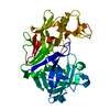
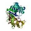
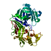
- Structure visualization
| Structure viewer | Molecule: MolmilJmol/JSmol |
|---|
- Downloads & links
-Download
| PDBx/mmCIF format | 1o5l.cif.gz | 42.9 KB | Display | PDBx/mmCIF format |
|---|---|---|---|---|
| PDB format | pdb1o5l.ent.gz | 29.4 KB | Display | PDB format |
| PDBx/mmJSON format | 1o5l.json.gz | Tree view | PDBx/mmJSON format | |
| Others |  Other downloads Other downloads |
-Validation report
| Arichive directory | https://data.pdbj.org/pub/pdb/validation_reports/o5/1o5lftp://data.pdbj.org/pub/pdb/validation_reports/o5/1o5l | HTTPS FTP |
|---|
-Related structure data


-Links
 PDBj
PDBj


- Assembly
| Deposited unit | 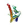
| ||||||||
|---|---|---|---|---|---|---|---|---|---|
| 1 | 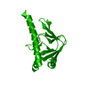
| ||||||||
| Unit cell |
|
-Components
| #1: Protein | Mass: 24895.195 Da / Num. of mol.: 1 Source method: isolated from a genetically manipulated source Source: (gene. exp.) Thermotoga maritima (bacteria) / Gene: TM1171 / Production host: |
|---|---|
| #2: Water | ChemComp-HOH /  Mass: 18.015 Da / Num. of mol.: 111 / Source method: isolated from a natural source / Formula: H2O Mass: 18.015 Da / Num. of mol.: 111 / Source method: isolated from a natural source / Formula: H2O |
-Experimental details
-Experiment
| Experiment | Method: X-RAY DIFFRACTION / Number of used crystals: 1 |
|---|
- Sample preparation
| Crystal | Density Matthews: 3.01 Å3/Da / Density % sol: 58.77 % |
|---|---|
| Crystal grow | Temperature: 277 K / Method: vapor diffusion, sitting drop, nanodrop / pH: 9.5 Details: 10% PEG-3000, 0.1M CHES pH 9.5, VAPOR DIFFUSION,SITTING DROP,NANODROP, temperature 277K |
| Crystal grow | *PLUS Temperature: 4 ℃ / pH: 5.5 / Method: vapor diffusion, sitting drop |
| Components of the solutions | *PLUS Conc.: 2.0 M / Common name: ammonium sulfate / Details: pH5.5 |
-Data collection
| Diffraction | Mean temperature: 100 K |
|---|---|
| Diffraction source | Source: SYNCHROTRON / Site: ALS  / Beamline: 5.0.2 / Wavelength: 0.9785 / Beamline: 5.0.2 / Wavelength: 0.9785 |
| Detector | Type: ADSC / Detector: CCD / Date: Jun 19, 2003 |
| Radiation | Monochromator: Double-crystal Si(111) / Protocol: SINGLE WAVELENGTH / Monochromatic (M) / Laue (L): M / Scattering type: x-ray |
| Radiation wavelength | Wavelength: 0.9785 Å / Relative weight: 1 |
| Reflection | Resolution: 2.05→50 Å / Num. all: 12634 / Num. obs: 12634 / % possible obs: 100 % / Redundancy: 5.82 % / Biso Wilson estimate: 53.02 Å2 / Rsym value: 0.112 / Net I/σ(I): 22.26 |
| Reflection shell | Resolution: 2.05→2.12 Å / Redundancy: 5.04 % / Mean I/σ(I) obs: 1.53 / Num. unique all: 1231 / Rsym value: 0.912 / % possible all: 98.17 |
| Reflection | *PLUS Highest resolution: 2.3 Å / Lowest resolution: 50 Å / Num. obs: 9367 / % possible obs: 99.4 % / Num. measured all: 73376 / Rmerge(I) obs: 0.082 |
| Reflection shell | *PLUS Highest resolution: 2.31 Å / Lowest resolution: 2.43 Å / % possible obs: 99.2 % / Rmerge(I) obs: 0.32 / Mean I/σ(I) obs: 4.2 |
- Processing
| Software |
| |||||||||||||||||||||||||||||||||||||||||||||||||||||||||||||||||||||||||||||||||||||||||||||||||||||||||||||||||||
|---|---|---|---|---|---|---|---|---|---|---|---|---|---|---|---|---|---|---|---|---|---|---|---|---|---|---|---|---|---|---|---|---|---|---|---|---|---|---|---|---|---|---|---|---|---|---|---|---|---|---|---|---|---|---|---|---|---|---|---|---|---|---|---|---|---|---|---|---|---|---|---|---|---|---|---|---|---|---|---|---|---|---|---|---|---|---|---|---|---|---|---|---|---|---|---|---|---|---|---|---|---|---|---|---|---|---|---|---|---|---|---|---|---|---|---|---|
| Refinement | Method to determine structure: SAD / Resolution: 2.3→38.97 Å / Cor.coef. Fo:Fc: 0.956 / Cor.coef. Fo:Fc free: 0.914 / SU B: 11.935 / SU ML: 0.154 / TLS residual ADP flag: LIKELY RESIDUAL / Isotropic thermal model: ISOTROPIC / Cross valid method: THROUGHOUT / ESU R: 0.239 / ESU R Free: 0.215 / Stereochemistry target values: MAXIMUM LIKELIHOOD Details: 1. HYDROGENS HAVE BEEN ADDED IN THE RIDING POSITIONS. 2. PROMINENT DENSITY NEAR RESIDUES SER64 AND ASP111 COULD BE RESIDUAL CAMP, SINCE THIS SEEMS TO BE A CAMP-BINDING CASSETTE.
| |||||||||||||||||||||||||||||||||||||||||||||||||||||||||||||||||||||||||||||||||||||||||||||||||||||||||||||||||||
| Solvent computation | Ion probe radii: 0.8 Å / Shrinkage radii: 0.8 Å / VDW probe radii: 1.2 Å / Solvent model: BABINET MODEL WITH MASK | |||||||||||||||||||||||||||||||||||||||||||||||||||||||||||||||||||||||||||||||||||||||||||||||||||||||||||||||||||
| Displacement parameters | Biso mean: 47.357 Å2
| |||||||||||||||||||||||||||||||||||||||||||||||||||||||||||||||||||||||||||||||||||||||||||||||||||||||||||||||||||
| Refinement step | Cycle: LAST / Resolution: 2.3→38.97 Å
| |||||||||||||||||||||||||||||||||||||||||||||||||||||||||||||||||||||||||||||||||||||||||||||||||||||||||||||||||||
| Refine LS restraints |
| |||||||||||||||||||||||||||||||||||||||||||||||||||||||||||||||||||||||||||||||||||||||||||||||||||||||||||||||||||
| LS refinement shell | Resolution: 2.3→2.36 Å / Total num. of bins used: 20
| |||||||||||||||||||||||||||||||||||||||||||||||||||||||||||||||||||||||||||||||||||||||||||||||||||||||||||||||||||
| Refinement TLS params. | Method: refined / Refine-ID: X-RAY DIFFRACTION
| |||||||||||||||||||||||||||||||||||||||||||||||||||||||||||||||||||||||||||||||||||||||||||||||||||||||||||||||||||
| Refinement TLS group | Refine-ID: X-RAY DIFFRACTION / Selection: ALL / Auth asym-ID: A / Label asym-ID: A
| |||||||||||||||||||||||||||||||||||||||||||||||||||||||||||||||||||||||||||||||||||||||||||||||||||||||||||||||||||
| Refinement | *PLUS Highest resolution: 2.3 Å / Lowest resolution: 50 Å / Rfactor Rfree: 0.253 / Rfactor Rwork: 0.197 | |||||||||||||||||||||||||||||||||||||||||||||||||||||||||||||||||||||||||||||||||||||||||||||||||||||||||||||||||||
| Solvent computation | *PLUS | |||||||||||||||||||||||||||||||||||||||||||||||||||||||||||||||||||||||||||||||||||||||||||||||||||||||||||||||||||
| Displacement parameters | *PLUS | |||||||||||||||||||||||||||||||||||||||||||||||||||||||||||||||||||||||||||||||||||||||||||||||||||||||||||||||||||
| Refine LS restraints | *PLUS
|
