Resolution: 1.65→35.93 Å / Cor.coef. Fo:Fc: 0.972 / Cor.coef. Fo:Fc free: 0.96 / SU B: 3.17 / SU ML: 0.053 / TLS residual ADP flag: LIKELY RESIDUAL / Isotropic thermal model: ISOTROPIC / Cross valid method: THROUGHOUT / ESU R: 0.078 / ESU R Free: 0.08 / Stereochemistry target values: MAXIMUM LIKELIHOOD Details: 1, RESIDUES A77-79 HAVE AMBIGUOUS DIFFERENCE DENSITY AROUND THEM AND MAY BE MISTRACED; RESIDUE A78 WAS OMITTED. RESIDUE A163 HAS AMBIGUOUS SIDECHAIN DENSITY. RESIDUES A147-148 MAY HAVE AN ...Details: 1, RESIDUES A77-79 HAVE AMBIGUOUS DIFFERENCE DENSITY AROUND THEM AND MAY BE MISTRACED; RESIDUE A78 WAS OMITTED. RESIDUE A163 HAS AMBIGUOUS SIDECHAIN DENSITY. RESIDUES A147-148 MAY HAVE AN ALTERNATIVE BACKBONE CONFORMATION, THAT WAS TOO AMBIGUOUS TO TRACE. THE ATOM 1 WAS BUILT AS CHLORINE BECAUSE IT PROVIDES THE BEST ELECTRONIC FIT FOR THE DIFFERENCE DENSITY, WHILE DISTANCES TO NEIGHBOURING WATERS ARE TOO LARGE FOR ISOELECTRONIC CATIONS. A POSSIBLE EXPLANATION FOR ITS PRESENCE IN THE UNCHARGED SITE IS THE LIKELY DIPOLE OF THE ADJACENT HELIX. 2, HYDROGENS HAVE BEEN ADDED IN THE RIDING POSITIONS.
Rfactor
Num. reflection
% reflection
Selection details
Rfree
0.18762
2036
5 %
RANDOM
Rwork
0.15768
-
-
-
obs
0.15918
38297
95.7 %
-
Solvent computation
Ion probe radii: 0.8 Å / Shrinkage radii: 0.8 Å / VDW probe radii: 1.4 Å / Solvent model: BABINET MODEL WITH MASK
Displacement parameters
Biso mean: 27.161 Å2
Baniso -1
Baniso -2
Baniso -3
1-
0.67 Å2
0 Å2
0 Å2
2-
-
1.31 Å2
0 Å2
3-
-
-
-1.98 Å2
Refinement step
Cycle: LAST / Resolution: 1.65→35.93 Å
Protein
Nucleic acid
Ligand
Solvent
Total
Num. atoms
2090
0
1
295
2386
Refine LS restraints
Refine-ID
Type
Dev ideal
Dev ideal target
Number
X-RAY DIFFRACTION
r_bond_refined_d
0.015
0.022
2155
X-RAY DIFFRACTION
r_bond_other_d
0.001
0.02
2004
X-RAY DIFFRACTION
r_angle_refined_deg
1.409
1.955
2912
X-RAY DIFFRACTION
r_angle_other_deg
0.755
3
4657
X-RAY DIFFRACTION
r_dihedral_angle_1_deg
5.544
5
263
X-RAY DIFFRACTION
r_dihedral_angle_2_deg
29.186
23.939
99
X-RAY DIFFRACTION
r_dihedral_angle_3_deg
14.127
15
392
X-RAY DIFFRACTION
r_dihedral_angle_4_deg
12.53
15
16
X-RAY DIFFRACTION
r_chiral_restr
0.083
0.2
326
X-RAY DIFFRACTION
r_gen_planes_refined
0.006
0.02
2365
X-RAY DIFFRACTION
r_gen_planes_other
0.001
0.02
431
X-RAY DIFFRACTION
r_nbd_refined
0.21
0.2
393
X-RAY DIFFRACTION
r_nbd_other
0.181
0.2
2008
X-RAY DIFFRACTION
r_nbtor_other
0.082
0.2
1268
X-RAY DIFFRACTION
r_xyhbond_nbd_refined
0.236
0.2
238
X-RAY DIFFRACTION
r_symmetry_vdw_refined
0.2
0.2
28
X-RAY DIFFRACTION
r_symmetry_vdw_other
0.245
0.2
119
X-RAY DIFFRACTION
r_symmetry_hbond_refined
0.26
0.2
40
X-RAY DIFFRACTION
r_mcbond_it
0.959
1.5
1321
X-RAY DIFFRACTION
r_mcangle_it
1.759
2
2142
X-RAY DIFFRACTION
r_scbond_it
2.522
3
834
X-RAY DIFFRACTION
r_scangle_it
4.32
4.5
770
LS refinement shell
Resolution: 1.65→1.693 Å / Total num. of bins used: 20
Rfactor
Num. reflection
% reflection
Rfree
0.276
121
5.38 %
Rwork
0.277
2128
-
Refinement TLS params.
Method: refined / Origin x: 8.823 Å / Origin y: 32.636 Å / Origin z: 20.851 Å
11
12
13
21
22
23
31
32
33
T
-0.1419 Å2
0.0143 Å2
-0.0017 Å2
-
-0.0695 Å2
-0.0001 Å2
-
-
-0.0558 Å2
L
0.1862 °2
0.2131 °2
-0.0032 °2
-
0.834 °2
-0.0158 °2
-
-
0.6055 °2
S
-0.0096 Å °
-0.0225 Å °
-0.0518 Å °
0.0403 Å °
0.027 Å °
-0.1607 Å °
0.065 Å °
0.0973 Å °
-0.0173 Å °
Refinement TLS group
Selection: ALL
+
About Yorodumi
-
News
-
Feb 9, 2022. New format data for meta-information of EMDB entries
New format data for meta-information of EMDB entries
Version 3 of the EMDB header file is now the official format.
The previous official version 1.9 will be removed from the archive.
In the structure databanks used in Yorodumi, some data are registered as the other names, "COVID-19 virus" and "2019-nCoV". Here are the details of the virus and the list of structure data.
Jan 31, 2019. EMDB accession codes are about to change! (news from PDBe EMDB page)
EMDB accession codes are about to change! (news from PDBe EMDB page)
The allocation of 4 digits for EMDB accession codes will soon come to an end. Whilst these codes will remain in use, new EMDB accession codes will include an additional digit and will expand incrementally as the available range of codes is exhausted. The current 4-digit format prefixed with “EMD-” (i.e. EMD-XXXX) will advance to a 5-digit format (i.e. EMD-XXXXX), and so on. It is currently estimated that the 4-digit codes will be depleted around Spring 2019, at which point the 5-digit format will come into force.
The EM Navigator/Yorodumi systems omit the EMD- prefix.
Related info.:Q: What is EMD? / ID/Accession-code notation in Yorodumi/EM Navigator
Yorodumi is a browser for structure data from EMDB, PDB, SASBDB, etc.
This page is also the successor to EM Navigator detail page, and also detail information page/front-end page for Omokage search.
The word "yorodu" (or yorozu) is an old Japanese word meaning "ten thousand". "mi" (miru) is to see.
Related info.:EMDB / PDB / SASBDB / Comparison of 3 databanks / Yorodumi Search / Aug 31, 2016. New EM Navigator & Yorodumi / Yorodumi Papers / Jmol/JSmol / Function and homology information / Changes in new EM Navigator and Yorodumi
 Movie
Movie Controller

 Yorodumi
 Open data
 Basic information
 Components
 Keywords
 Function and homology information
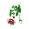
 Thermotoga maritima (bacteria)
 X-RAY DIFFRACTION /
  Authors
 Citation
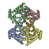
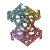
 Structure visualization
 Downloads & links
 Other downloads
 PDBj
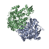
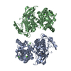
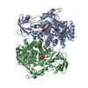
PDBj Assembly

 Mass: 35.453 Da / Num. of mol.: 1 / Source method: obtained synthetically / Formula: Cl
 Mass: 18.015 Da / Num. of mol.: 295 / Source method: isolated from a natural source / Formula: H2O
 Sample preparation
 / Beamline: BL11-1 / Wavelength: 0.976224
  Processing