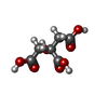
+ Open data
- Basic information
| Entry | Database: PDB / ID: 1o4l | ||||||
|---|---|---|---|---|---|---|---|
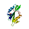
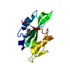
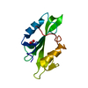
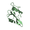
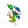
| Title | CRYSTAL STRUCTURE OF SH2 IN COMPLEX WITH FRAGMENT2. | ||||||
 Components Components | PROTO-ONCOGENE TYROSINE-PROTEIN KINASE SRC | ||||||
 Keywords Keywords | SIGNALING PROTEIN / SH2 DOMAIN FRAGMENT APPROACH | ||||||
| Function / homology |  Function and homology information Function and homology informationregulation of caveolin-mediated endocytosis / regulation of toll-like receptor 3 signaling pathway / cellular response to progesterone stimulus / positive regulation of platelet-derived growth factor receptor-beta signaling pathway / positive regulation of dephosphorylation / regulation of cell projection assembly / negative regulation of telomere maintenance / Regulation of commissural axon pathfinding by SLIT and ROBO / regulation of epithelial cell migration / ERBB2 signaling pathway ...regulation of caveolin-mediated endocytosis / regulation of toll-like receptor 3 signaling pathway / cellular response to progesterone stimulus / positive regulation of platelet-derived growth factor receptor-beta signaling pathway / positive regulation of dephosphorylation / regulation of cell projection assembly / negative regulation of telomere maintenance / Regulation of commissural axon pathfinding by SLIT and ROBO / regulation of epithelial cell migration / ERBB2 signaling pathway / Regulation of gap junction activity / negative regulation of focal adhesion assembly / BMP receptor binding / positive regulation of integrin activation / positive regulation of protein processing / Activated NTRK2 signals through FYN / Netrin mediated repulsion signals / regulation of intracellular estrogen receptor signaling pathway / intestinal epithelial cell development / negative regulation of neutrophil activation / regulation of vascular permeability / focal adhesion assembly / connexin binding / osteoclast development / Activated NTRK3 signals through PI3K / cellular response to fluid shear stress / signal complex assembly / positive regulation of small GTPase mediated signal transduction / branching involved in mammary gland duct morphogenesis / Co-stimulation by CD28 / Regulation of RUNX1 Expression and Activity / DCC mediated attractive signaling / EPH-Ephrin signaling / positive regulation of podosome assembly / positive regulation of lamellipodium morphogenesis / regulation of bone resorption / Ephrin signaling / Signal regulatory protein family interactions / odontogenesis / negative regulation of mitochondrial depolarization / podosome / MET activates PTK2 signaling / cellular response to peptide hormone stimulus / Regulation of KIT signaling / regulation of early endosome to late endosome transport / Signaling by ALK / leukocyte migration / phospholipase activator activity / oogenesis / Co-inhibition by CTLA4 / GP1b-IX-V activation signalling / EPHA-mediated growth cone collapse / Receptor Mediated Mitophagy / p130Cas linkage to MAPK signaling for integrins / interleukin-6-mediated signaling pathway / stress fiber assembly / positive regulation of Notch signaling pathway / Signaling by EGFR / RUNX2 regulates osteoblast differentiation / stimulatory C-type lectin receptor signaling pathway / negative regulation of intrinsic apoptotic signaling pathway / Fc-gamma receptor signaling pathway involved in phagocytosis / forebrain development / regulation of cell-cell adhesion / uterus development / PECAM1 interactions / Recycling pathway of L1 / GRB2:SOS provides linkage to MAPK signaling for Integrins / regulation of heart rate by cardiac conduction / RHOU GTPase cycle / protein tyrosine kinase activator activity / RET signaling / signaling receptor activator activity / negative regulation of anoikis / FCGR activation / Long-term potentiation / positive regulation of epithelial cell migration / progesterone receptor signaling pathway / positive regulation of protein serine/threonine kinase activity / EPH-ephrin mediated repulsion of cells / GAB1 signalosome / ephrin receptor signaling pathway / vascular endothelial growth factor receptor signaling pathway / negative regulation of hippo signaling / bone resorption / negative regulation of protein-containing complex assembly / Nuclear signaling by ERBB4 / phospholipase binding / ephrin receptor binding / T cell costimulation / cellular response to platelet-derived growth factor stimulus / p38MAPK events / Signaling by ERBB2 / Integrin signaling / EPHB-mediated forward signaling / ionotropic glutamate receptor binding / positive regulation of TORC1 signaling / NCAM signaling for neurite out-growth / Downregulation of ERBB4 signaling / Downstream signal transduction Similarity search - Function | ||||||
| Biological species |  Homo sapiens (human) Homo sapiens (human) | ||||||
| Method |  X-RAY DIFFRACTION / X-RAY DIFFRACTION /  molecular replacement / Resolution: 1.65 Å molecular replacement / Resolution: 1.65 Å | ||||||
 Authors Authors | Lange, G. / Loenze, P. / Liesum, A. | ||||||
 Citation Citation |  Journal: J.Med.Chem. / Year: 2003 Journal: J.Med.Chem. / Year: 2003Title: Requirements for specific binding of low affinity inhibitor fragments to the SH2 domain of (pp60)Src are identical to those for high affinity binding of full length inhibitors. Authors: Lange, G. / Lesuisse, D. / Deprez, P. / Schoot, B. / Loenze, P. / Benard, D. / Marquette, J.P. / Broto, P. / Sarubbi, E. / Mandine, E. | ||||||
| History |
|
- Structure visualization
| Structure viewer | Molecule:  Molmil Molmil Jmol/JSmol Jmol/JSmol |
|---|
- Downloads & links
- Download
| PDBx/mmCIF format |  1o4l.cif.gz 1o4l.cif.gz | 36.9 KB | Display |  PDBx/mmCIF format PDBx/mmCIF format |
|---|---|---|---|---|
| PDB format |  pdb1o4l.ent.gz pdb1o4l.ent.gz | 24.7 KB | Display |  PDB format PDB format |
| PDBx/mmJSON format |  1o4l.json.gz 1o4l.json.gz | Tree view |  PDBx/mmJSON format PDBx/mmJSON format | |
| Others |  Other downloads Other downloads |
-Validation report
| Summary document |  1o4l_validation.pdf.gz 1o4l_validation.pdf.gz | 387.2 KB | Display |  wwPDB validaton report wwPDB validaton report |
|---|---|---|---|---|
| Full document |  1o4l_full_validation.pdf.gz 1o4l_full_validation.pdf.gz | 388.3 KB | Display | |
| Data in XML |  1o4l_validation.xml.gz 1o4l_validation.xml.gz | 4.2 KB | Display | |
| Data in CIF |  1o4l_validation.cif.gz 1o4l_validation.cif.gz | 6.7 KB | Display | |
| Arichive directory |  https://data.pdbj.org/pub/pdb/validation_reports/o4/1o4l https://data.pdbj.org/pub/pdb/validation_reports/o4/1o4l ftp://data.pdbj.org/pub/pdb/validation_reports/o4/1o4l ftp://data.pdbj.org/pub/pdb/validation_reports/o4/1o4l | HTTPS FTP |
-Related structure data
| Related structure data |  1o41C  1o42C  1o43C  1o44C  1o45C 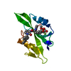 1o46C  1o47C  1o48C  1o49C  1o4aC  1o4bC  1o4cC  1o4dC  1o4eC  1o4fC  1o4gC  1o4hC  1o4iC  1o4jC  1o4kC  1o4mC  1o4nC  1o4oC  1o4pC  1o4qC  1o4rC  1shdS S: Starting model for refinement C: citing same article ( |
|---|---|
| Similar structure data |
- Links
Links
- Assembly
| Deposited unit | 
| ||||||||
|---|---|---|---|---|---|---|---|---|---|
| 1 |
| ||||||||
| Unit cell |
|
- Components
| #1: Protein | Mass: 12374.964 Da / Num. of mol.: 1 / Fragment: SH2 DOMAIN Source method: isolated from a genetically manipulated source Source: (gene. exp.)  Homo sapiens (human) / Gene: SRC / Plasmid: BL21 (DE3) / Production host: Homo sapiens (human) / Gene: SRC / Plasmid: BL21 (DE3) / Production host:  |
|---|---|
| #2: Chemical | ChemComp-CIT / |
| #3: Water | ChemComp-HOH / |
-Experimental details
-Experiment
| Experiment | Method:  X-RAY DIFFRACTION / Number of used crystals: 1 X-RAY DIFFRACTION / Number of used crystals: 1 |
|---|
- Sample preparation
| Crystal | Density Matthews: 2.2 Å3/Da / Density % sol: 41.9 % | ||||||||||||||||||||||||||||||||||||||||||
|---|---|---|---|---|---|---|---|---|---|---|---|---|---|---|---|---|---|---|---|---|---|---|---|---|---|---|---|---|---|---|---|---|---|---|---|---|---|---|---|---|---|---|---|
| Crystal grow | pH: 5.5 / Details: pH 5.50 | ||||||||||||||||||||||||||||||||||||||||||
| Crystal grow | *PLUS pH: 5.5 / Method: vapor diffusion, sitting drop / Details: Lesuisse, D., (2002) J.Med.Chem., 45, 2379. | ||||||||||||||||||||||||||||||||||||||||||
| Components of the solutions | *PLUS
|
-Data collection
| Diffraction | Mean temperature: 100 K |
|---|---|
| Diffraction source | Source:  ROTATING ANODE / Type: ELLIOTT GX-21 / Wavelength: 1.5418 ROTATING ANODE / Type: ELLIOTT GX-21 / Wavelength: 1.5418 |
| Detector | Type: MAR scanner 345 mm plate / Detector: IMAGE PLATE / Date: Nov 5, 1997 |
| Radiation | Monochromator: GRAPHITE / Protocol: SINGLE WAVELENGTH / Monochromatic (M) / Laue (L): M / Scattering type: x-ray |
| Radiation wavelength | Wavelength: 1.5418 Å / Relative weight: 1 |
| Reflection | Resolution: 1.65→40 Å / Num. obs: 12473 / % possible obs: 98.9 % / Observed criterion σ(I): -3 / Rmerge(I) obs: 0.057 |
- Processing
| Software |
| ||||||||||||||||||||||||||||||||||||||||||||||||||||||||||||
|---|---|---|---|---|---|---|---|---|---|---|---|---|---|---|---|---|---|---|---|---|---|---|---|---|---|---|---|---|---|---|---|---|---|---|---|---|---|---|---|---|---|---|---|---|---|---|---|---|---|---|---|---|---|---|---|---|---|---|---|---|---|
| Refinement | Method to determine structure:  molecular replacement molecular replacementStarting model: 1SHD Resolution: 1.65→8 Å / Data cutoff high absF: 1000000 / Data cutoff low absF: 0.1
| ||||||||||||||||||||||||||||||||||||||||||||||||||||||||||||
| Displacement parameters | Biso mean: 23.9 Å2 | ||||||||||||||||||||||||||||||||||||||||||||||||||||||||||||
| Refinement step | Cycle: LAST / Resolution: 1.65→8 Å
| ||||||||||||||||||||||||||||||||||||||||||||||||||||||||||||
| Refine LS restraints |
|
 Movie
Movie Controller





 PDBj
PDBj


