 Movie
Movie Controller

[English] 日本語
 Yorodumi
- PDB-1o2a: Crystal structure of Thymidylate Synthase Complementing Protein (... -
+ Open data

- Basic information
| Entry | Database: PDB / ID: 1o2a | ||||||
|---|---|---|---|---|---|---|---|
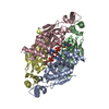
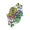
| Title | Crystal structure of Thymidylate Synthase Complementing Protein (TM0449) from Thermotoga maritima with FAD at 1.8 A resolution | ||||||
 Components Components | Thymidylate synthase thyX | ||||||
 Keywords Keywords | TRANSFERASE / TM0449 / THYMIDYLATE SYNTHASE COMPLEMENTING PROTEIN / STRUCTURAL GENOMICS / JCSG / Joint Center for Structural Genomics / PSI / Protein Structure Initiative | ||||||
| Function / homology |  Function and homology information Function and homology informationthymidylate synthase (FAD) / thymidylate synthase (FAD) activity / thymidylate synthase activity / dTMP biosynthetic process / dTTP biosynthetic process / NADPH binding / flavin adenine dinucleotide binding / methylation Similarity search - Function | ||||||
| Biological species |   Thermotoga maritima (bacteria) Thermotoga maritima (bacteria) | ||||||
| Method |  X-RAY DIFFRACTION / SYNCHROTRON / MOLECULAR REPLACEMENT / Resolution: 1.8 Å X-RAY DIFFRACTION / SYNCHROTRON / MOLECULAR REPLACEMENT / Resolution: 1.8 Å | ||||||
 Authors Authors | Mathews, I.I. / Deacon, A.M. / Canaves, J.M. / McMullan, D. / Lesley, S.A. / Agarwalla, S. / Kuhn, P. / Joint Center for Structural Genomics (JCSG) | ||||||
 Citation Citation | Journal: Structure / Year: 2003 Title: Functional Analysis of Substrate and Cofactor Complex Structures of a Thymidylate Synthase-Complementing Protein Authors: Mathews, I.I. / Deacon, A.M. / Canaves, J.M. / McMullan, D. / Lesley, S.A. / Agarwalla, S. / Kuhn, P. | ||||||
| History |
|
- Structure visualization
| Structure viewer | Molecule: MolmilJmol/JSmol |
|---|
- Downloads & links
-Download
| PDBx/mmCIF format | 1o2a.cif.gz | 197.4 KB | Display | PDBx/mmCIF format |
|---|---|---|---|---|
| PDB format | pdb1o2a.ent.gz | 158.8 KB | Display | PDB format |
| PDBx/mmJSON format | 1o2a.json.gz | Tree view | PDBx/mmJSON format | |
| Others |  Other downloads Other downloads |
-Validation report
| Summary document | 1o2a_validation.pdf.gz | 1.6 MB | Display | wwPDB validaton report |
|---|---|---|---|---|
| Full document | 1o2a_full_validation.pdf.gz | 1.7 MB | Display | |
| Data in XML | 1o2a_validation.xml.gz | 40.7 KB | Display | |
| Data in CIF | 1o2a_validation.cif.gz | 52.7 KB | Display | |
| Arichive directory | https://data.pdbj.org/pub/pdb/validation_reports/o2/1o2aftp://data.pdbj.org/pub/pdb/validation_reports/o2/1o2a | HTTPS FTP |
-Related structure data
| Related structure data |  1o24C 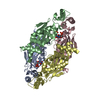 1o25C 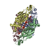 1o26C  1o27C 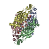 1o28C  1o29C  1o2bC  1kq4S S: Starting model for refinement C: citing same article ( |
|---|---|
| Similar structure data |


-Links
 PDBj
PDBj- Assembly
| Deposited unit | 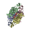
| ||||||||
|---|---|---|---|---|---|---|---|---|---|
| 1 |
| ||||||||
| Unit cell |
|
-Components
| #1: Protein | Mass: 27503.680 Da / Num. of mol.: 4 Source method: isolated from a genetically manipulated source Source: (gene. exp.) Thermotoga maritima (bacteria) / Gene: TM0449 / Production host: References: UniProt: Q9WYT0, Transferases; Transferring one-carbon groups; Methyltransferases #2: Chemical | ChemComp-FAD /   Mass: 785.550 Da / Num. of mol.: 4 / Source method: obtained synthetically / Formula: C27H33N9O15P2 / Comment: FAD*YM Mass: 785.550 Da / Num. of mol.: 4 / Source method: obtained synthetically / Formula: C27H33N9O15P2 / Comment: FAD*YM#3: Water | ChemComp-HOH / |  Mass: 18.015 Da / Num. of mol.: 308 / Source method: isolated from a natural source / Formula: H2O Mass: 18.015 Da / Num. of mol.: 308 / Source method: isolated from a natural source / Formula: H2O |
|---|
-Experimental details
-Experiment
| Experiment | Method: X-RAY DIFFRACTION / Number of used crystals: 1 |
|---|
- Sample preparation
| Crystal | Density Matthews: 2.1 Å3/Da / Density % sol: 41 % | ||||||||||||||||||||||||
|---|---|---|---|---|---|---|---|---|---|---|---|---|---|---|---|---|---|---|---|---|---|---|---|---|---|
| Crystal grow | Temperature: 295 K / Method: vapor diffusion, hanging drop / pH: 8 Details: 49% PEG 200, 0.1M Tris-HCL, pH 8.0, VAPOR DIFFUSION,HANGING DROP, temperature 295K | ||||||||||||||||||||||||
| Crystal grow | *PLUS pH: 7.5 / Method: vapor diffusion, sitting drop / Details: Kuhn, P., (2002) Proteins, 49, 142. | ||||||||||||||||||||||||
| Components of the solutions | *PLUS
|
-Data collection
| Diffraction | Mean temperature: 100 K |
|---|---|
| Diffraction source | Source: SYNCHROTRON / Site: SSRL  / Beamline: BL11-1 / Wavelength: 0.98008 / Beamline: BL11-1 / Wavelength: 0.98008 |
| Detector | Type: ADSC QUANTUM 315 / Detector: CCD / Date: May 24, 2002 Details: Flat mirror, single crystal Si(111) bent monochromator |
| Radiation | Protocol: SINGLE WAVELENGTH / Monochromatic (M) / Laue (L): M / Scattering type: x-ray |
| Radiation wavelength | Wavelength: 0.98008 Å / Relative weight: 1 |
| Reflection | Resolution: 1.7→46.641 Å / Num. all: 86542 / Num. obs: 86542 / % possible obs: 86.7 % / Redundancy: 3.4 % / Biso Wilson estimate: 25.613 Å2 / Rsym value: 0.034 / Net I/σ(I): 19.4 |
| Reflection shell | Resolution: 1.7→1.74 Å / Redundancy: 2.9 % / Mean I/σ(I) obs: 2.8 / Num. unique all: 4020 / Rsym value: 0.49 / % possible all: 56.2 |
| Reflection | *PLUS Highest resolution: 1.8 Å / Lowest resolution: 38.3 Å / Num. obs: 78182 / % possible obs: 92.5 % / Num. measured all: 331090 / Rmerge(I) obs: 0.045 |
| Reflection shell | *PLUS % possible obs: 81.4 % / Rmerge(I) obs: 0.239 |
- Processing
| Software |
| ||||||||||||||||||||||||||||
|---|---|---|---|---|---|---|---|---|---|---|---|---|---|---|---|---|---|---|---|---|---|---|---|---|---|---|---|---|---|
| Refinement | Method to determine structure: MOLECULAR REPLACEMENT Starting model: 1KQ4 Resolution: 1.8→20 Å / Isotropic thermal model: Isotropic / Cross valid method: THROUGHOUT / σ(F): 2 / Stereochemistry target values: Engh and Huber Details: Alternate conformation B of residue 91 of chain D is not linked to the N of SER 92 chain D. Alternate conformation A is covalently linked.
| ||||||||||||||||||||||||||||
| Solvent computation | Solvent model: Bulk solvent correction / Bsol: 0.394268 Å2 / ksol: 50.7167 e/Å3 | ||||||||||||||||||||||||||||
| Displacement parameters | Biso mean: 40.6 Å2
| ||||||||||||||||||||||||||||
| Refine analyze | Luzzati coordinate error obs: 0.2041 Å | ||||||||||||||||||||||||||||
| Refinement step | Cycle: LAST / Resolution: 1.8→20 Å
| ||||||||||||||||||||||||||||
| Refine LS restraints |
| ||||||||||||||||||||||||||||
| LS refinement shell | Resolution: 1.8→1.81 Å / Total num. of bins used: 50
| ||||||||||||||||||||||||||||
| Refinement | *PLUS Highest resolution: 1.8 Å / Lowest resolution: 20 Å / Rfactor Rfree: 0.23 / Rfactor Rwork: 0.203 / % reflection Rfree: 5 % | ||||||||||||||||||||||||||||
| Solvent computation | *PLUS | ||||||||||||||||||||||||||||
| Displacement parameters | *PLUS |
