 Movie
Movie Controller

[English] 日本語
 Yorodumi
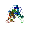
- PDB-1nwd: Solution Structure of Ca2+/Calmodulin bound to the C-terminal Dom... -
+ Open data

- Basic information
| Entry | Database: PDB / ID: 1nwd | ||||||
|---|---|---|---|---|---|---|---|
| Title | Solution Structure of Ca2+/Calmodulin bound to the C-terminal Domain of Petunia Glutamate Decarboxylase | ||||||
 Components Components |
| ||||||
 Keywords Keywords | BINDING PROTEIN/HYDROLASE / CALMODULIN-PEPTIDE COMPLEX / CALMODULIN / GAD / GLUTAMATE DECARBOXYLASE / DIMER / BINDING PROTEIN-HYDROLASE COMPLEX | ||||||
| Function / homology |  Function and homology information Function and homology informationglutamate decarboxylase / glutamate decarboxylase activity / L-glutamate catabolic process / myosin II complex / pyridoxal phosphate binding / calmodulin binding / signaling receptor binding / calcium ion binding / cytosol Similarity search - Function | ||||||
| Biological species |  Petunia x hybrida (garden petunia) Petunia x hybrida (garden petunia) | ||||||
| Method | SOLUTION NMR / simulated annealing, torsion angle dynamics | ||||||
 Authors Authors | Yap, K.L. / Yuan, T. / Mal, T.K. / Vogel, H.J. / Ikura, M. | ||||||
 Citation Citation | Journal: J.Mol.Biol. / Year: 2003 Title: Structural Basis for Simultaneous Binding of Two Carboxy-terminal Peptides of Plant Glutamate Decarboxylase to Calmodulin Authors: Yap, K.L. / Yuan, T. / Mal, T.K. / Vogel, H.J. / Ikura, M. #1: Journal: J.Biol.Chem. / Year: 1998Title: Calcium-calmodulin-induced dimerization of the carboxyl-terminal domain from Petunia glutamate decarboxylase | ||||||
| History |
| ||||||
| Remark 999 | SEQUENCE The scientific organism for Calmodulin in this entry is Xenopus leavis (African clawed ...SEQUENCE The scientific organism for Calmodulin in this entry is Xenopus leavis (African clawed frog) and the sequence is identical to that of the human Calmodulin (Swiss-Prot accesssion CALM_HUMAN P02593). There is no separate sequence for Xenopus laevis and because it is 100% identical to that of human, Swiss-Prot also lists Xenopus laevis as a source for this sequence accession number. |
- Structure visualization
| Structure viewer | Molecule: MolmilJmol/JSmol |
|---|
- Downloads & links
-Download
| PDBx/mmCIF format | 1nwd.cif.gz | 1.2 MB | Display | PDBx/mmCIF format |
|---|---|---|---|---|
| PDB format | pdb1nwd.ent.gz | 1 MB | Display | PDB format |
| PDBx/mmJSON format | 1nwd.json.gz | Tree view | PDBx/mmJSON format | |
| Others |  Other downloads Other downloads |
-Validation report
| Arichive directory | https://data.pdbj.org/pub/pdb/validation_reports/nw/1nwdftp://data.pdbj.org/pub/pdb/validation_reports/nw/1nwd | HTTPS FTP |
|---|
-Related structure data
| Similar structure data |
|---|
-Links
 PDBj
PDBj
- Assembly
| Deposited unit | 
| |||||||||
|---|---|---|---|---|---|---|---|---|---|---|
| 1 |
| |||||||||
| NMR ensembles |
|
-Components
| #1: Protein | Mass: 16721.350 Da / Num. of mol.: 1 Source method: isolated from a genetically manipulated source Source: (gene. exp.) Gene: (CALM1 OR CAM1 OR CALM OR CAM) AND (CALM2 OR CAM2 OR CAMB) AND (CALM3 OR CAM3 OR CAMC) Plasmid: pAS / Production host:  | ||
|---|---|---|---|
| #2: Protein/peptide | Mass: 3342.922 Da / Num. of mol.: 2 Fragment: C-TERMINAL CALMODULIN BINDING DOMAIN (residues 470-495) Source method: isolated from a genetically manipulated source Source: (gene. exp.) Petunia x hybrida (garden petunia) / Gene: GAD / Plasmid: pGEX-2T / Species (production host): Escherichia coli / Production host: #3: Chemical | ChemComp-CA /   Mass: 40.078 Da / Num. of mol.: 4 / Source method: obtained synthetically / Formula: Ca Mass: 40.078 Da / Num. of mol.: 4 / Source method: obtained synthetically / Formula: Ca |
-Experimental details
-Experiment
| Experiment | Method: SOLUTION NMR |
|---|---|
| NMR details | Text: This structure was determined using isotope-edited and filtered triple resonance NMR experiments on a number of complex samples with various combinations of 15-N, 13-C and 2-H labeling. See ...Text: This structure was determined using isotope-edited and filtered triple resonance NMR experiments on a number of complex samples with various combinations of 15-N, 13-C and 2-H labeling. See Citation reference for details. |
- Sample preparation
| Details | Contents: 1 mM Calmodulin; 2.2 mM GAD / Solvent system: 91% H2O, 9% D2O |
|---|---|
| Sample conditions | Ionic strength: 100 mM KCl, 10 mM CaCl2 / pH: 6.3 / Temperature: 308 K |
| Crystal grow | *PLUS Method: other / Details: NMR |
-NMR measurement
| Radiation | Protocol: SINGLE WAVELENGTH / Monochromatic (M) / Laue (L): M | |||||||||||||||
|---|---|---|---|---|---|---|---|---|---|---|---|---|---|---|---|---|
| Radiation wavelength | Relative weight: 1 | |||||||||||||||
| NMR spectrometer |
|
- Processing
| NMR software |
| ||||||||||||||||||||
|---|---|---|---|---|---|---|---|---|---|---|---|---|---|---|---|---|---|---|---|---|---|
| Refinement | Method: simulated annealing, torsion angle dynamics / Software ordinal: 1 | ||||||||||||||||||||
| NMR representative | Selection criteria: lowest energy | ||||||||||||||||||||
| NMR ensemble | Conformer selection criteria: structures with the lowest energy Conformers calculated total number: 200 / Conformers submitted total number: 20 |
 NMRPipe