 Movie
Movie Controller

[English] 日本語
 Yorodumi
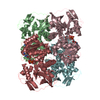
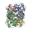
- PDB-1nto: N249Y MUTANT OF ALCOHOL DEHYDROGENASE FROM THE ARCHAEON SULFOLOBU... -
+ Open data

- Basic information
| Entry | Database: PDB / ID: 1nto | ||||||
|---|---|---|---|---|---|---|---|
| Title | N249Y MUTANT OF ALCOHOL DEHYDROGENASE FROM THE ARCHAEON SULFOLOBUS SOLFATARICUS-MONOCLINIC CRYSTAL FORM | ||||||
 Components Components | NAD-dependent alcohol dehydrogenase | ||||||
 Keywords Keywords | OXIDOREDUCTASE / ARCHAEON / NAD(H)-DEPENDENT / MUTANT | ||||||
| Function / homology |  Function and homology information Function and homology informationalcohol dehydrogenase (NAD+) activity / alcohol dehydrogenase / oxidoreductase activity / zinc ion binding Similarity search - Function | ||||||
| Biological species |   Sulfolobus solfataricus (archaea) Sulfolobus solfataricus (archaea) | ||||||
| Method |  X-RAY DIFFRACTION / SYNCHROTRON / MOLECULAR REPLACEMENT / Resolution: 1.94 Å X-RAY DIFFRACTION / SYNCHROTRON / MOLECULAR REPLACEMENT / Resolution: 1.94 Å | ||||||
 Authors Authors | Esposito, L. / Bruno, I. / Sica, F. / Raia, C.A. / Giordano, A. / Rossi, M. / Mazzarella, L. / Zagari, A. | ||||||
 Citation Citation | Journal: Febs Lett. / Year: 2003 Title: Structural study of a single-point mutant of Sulfolobus solfataricus alcohol dehydrogenase with enhanced activity. Authors: Esposito, L. / Bruno, I. / Sica, F. / Raia, C.A. / Giordano, A. / Rossi, M. / Mazzarella, L. / Zagari, A. #1: Journal: Biochemistry / Year: 1999Title: Asn249tyr substitution at the coenzyme binding domain activates Sulfolobus solfataricus alcohol dehydrogenase and increases its thermal stability Authors: Giordano, A. / Cannio, R. / La Cara, F. / Bartolucci, S. / Rossi, M. / Raia, C.A. #2: Journal: J.Mol.Biol. / Year: 2002Title: Crystal structure of alcohol dehydrogenase from the hyperthermophilic archaeon Sulfolobus solfataricus at 1.85 angstrom resolution Authors: Esposito, L. / Sica, F. / Raia, C.A. / Giordano, A. / Rossi, M. / Mazzarella, L. / Zagari, A. #3: Journal: J.Mol.Biol. / Year: 1976Title: Three Dimensional Structure of Horse Liver Alcoholdehydrogenase at 2.4 Angstrom Resolution Authors: Eklund, H. / Nordstrom, B. / Zeppezauer, E. / Soderlund, G. / Ohlsson, I. / Boiwe, T. / Soderberg, B.O. / Tapia, O. / Branden, C.I. / Akeson, A. | ||||||
| History |
|
- Structure visualization
| Structure viewer | Molecule: MolmilJmol/JSmol |
|---|
- Downloads & links
-Download
| PDBx/mmCIF format | 1nto.cif.gz | 401.8 KB | Display | PDBx/mmCIF format |
|---|---|---|---|---|
| PDB format | pdb1nto.ent.gz | 329.4 KB | Display | PDB format |
| PDBx/mmJSON format | 1nto.json.gz | Tree view | PDBx/mmJSON format | |
| Others |  Other downloads Other downloads |
-Validation report
| Arichive directory | https://data.pdbj.org/pub/pdb/validation_reports/nt/1ntoftp://data.pdbj.org/pub/pdb/validation_reports/nt/1nto | HTTPS FTP |
|---|
-Related structure data
| Related structure data |  1nvgC  1jvbS S: Starting model for refinement C: citing same article ( |
|---|---|
| Similar structure data |


-Links
 PDBj
PDBj

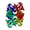
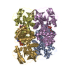
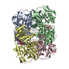
- Assembly
| Deposited unit | 
| ||||||||
|---|---|---|---|---|---|---|---|---|---|
| 1 | 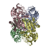
| ||||||||
| 2 | 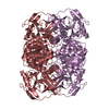
| ||||||||
| Unit cell |
| ||||||||
| Details | The biological assembly is a tetramer. The asymmetric unit contains six monomers. Chains ABCD form a tetramer. The second tetramer is generated from chains EH in the asymmetric unit by the operation: -x+1, y, -z+1 |
-Components
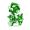
| #1: Protein | Mass: 37664.594 Da / Num. of mol.: 6 / Mutation: N249Y Source method: isolated from a genetically manipulated source Source: (gene. exp.) Sulfolobus solfataricus (archaea) / Gene: ADH / Plasmid: PTRC99A / Production host:  #2: Chemical | ChemComp-ZN /   Mass: 65.409 Da / Num. of mol.: 12 / Source method: obtained synthetically / Formula: Zn Mass: 65.409 Da / Num. of mol.: 12 / Source method: obtained synthetically / Formula: Zn#3: Water | ChemComp-HOH / |  Mass: 18.015 Da / Num. of mol.: 633 / Source method: isolated from a natural source / Formula: H2O Mass: 18.015 Da / Num. of mol.: 633 / Source method: isolated from a natural source / Formula: H2O |
|---|
-Experimental details
-Experiment
| Experiment | Method: X-RAY DIFFRACTION / Number of used crystals: 1 |
|---|
- Sample preparation
| Crystal | Density Matthews: 2.51 Å3/Da / Density % sol: 50.68 % |
|---|---|
| Crystal grow | Temperature: 296 K / Method: vapor diffusion, hanging drop Details: 7%(w/v) PEG 4000, 7%(v/v) Isopropanol, 50 mM Sodium citrate pH 5.6, 50 mM Tris pH 7.8 , 25 mM CoCl2 , VAPOR DIFFUSION, HANGING DROP, temperature 296K |
| Crystal grow | *PLUS Details: Pearl, L., (1993) J. Mol. Biol., 229, 782. |
-Data collection
| Diffraction | Mean temperature: 110 K |
|---|---|
| Diffraction source | Source: SYNCHROTRON / Site: ELETTRA  / Beamline: 5.2R / Wavelength: 1 / Wavelength: 1 Å / Beamline: 5.2R / Wavelength: 1 / Wavelength: 1 Å |
| Detector | Type: MARRESEARCH / Detector: IMAGE PLATE / Date: Jun 5, 1998 |
| Radiation | Protocol: SINGLE WAVELENGTH / Monochromatic (M) / Laue (L): M / Scattering type: x-ray |
| Radiation wavelength | Wavelength: 1 Å / Relative weight: 1 |
| Reflection | Resolution: 1.94→15 Å / Num. obs: 146709 / % possible obs: 87.9 % / Redundancy: 3.8 % / Rmerge(I) obs: 0.033 / Net I/σ(I): 29.8 |
| Reflection shell | Resolution: 1.94→2.01 Å / Redundancy: 2.8 % / Rmerge(I) obs: 0.09 / Mean I/σ(I) obs: 11.7 / % possible all: 86.9 |
| Reflection | *PLUS Lowest resolution: 15 Å / Num. measured all: 554277 |
| Reflection shell | *PLUS % possible obs: 86.9 % / Rmerge(I) obs: 0.09 / Mean I/σ(I) obs: 12 |
- Processing
| Software |
| ||||||||||||||||||||||||||||||||||||
|---|---|---|---|---|---|---|---|---|---|---|---|---|---|---|---|---|---|---|---|---|---|---|---|---|---|---|---|---|---|---|---|---|---|---|---|---|---|
| Refinement | Method to determine structure: MOLECULAR REPLACEMENT Starting model: PDB ENTRY 1JVB Resolution: 1.94→15 Å / Rfactor Rfree error: 0.003 / Isotropic thermal model: RESTRAINED / Cross valid method: THROUGHOUT / σ(F): 0 / Stereochemistry target values: Engh & Huber
| ||||||||||||||||||||||||||||||||||||
| Solvent computation | Solvent model: FLAT MODEL / Bsol: 61.59 Å2 / ksol: 0.4714 e/Å3 | ||||||||||||||||||||||||||||||||||||
| Displacement parameters | Biso mean: 36.5 Å2
| ||||||||||||||||||||||||||||||||||||
| Refine analyze |
| ||||||||||||||||||||||||||||||||||||
| Refinement step | Cycle: LAST / Resolution: 1.94→15 Å
| ||||||||||||||||||||||||||||||||||||
| Refine LS restraints |
| ||||||||||||||||||||||||||||||||||||
| LS refinement shell | Resolution: 1.94→2.01 Å / Rfactor Rfree error: 0.011 / Total num. of bins used: 10
| ||||||||||||||||||||||||||||||||||||
| Xplor file |
| ||||||||||||||||||||||||||||||||||||
| Refinement | *PLUS % reflection Rfree: 5 % / Rfactor Rwork: 0.21 | ||||||||||||||||||||||||||||||||||||
| Solvent computation | *PLUS | ||||||||||||||||||||||||||||||||||||
| Displacement parameters | *PLUS | ||||||||||||||||||||||||||||||||||||
| Refine LS restraints | *PLUS
|
