 Movie
Movie Controller

[English] 日本語
 Yorodumi
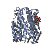
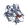
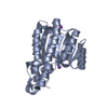
- PDB-1nrf: C-terminal domain of the Bacillus licheniformis BlaR penicillin-r... -
+ Open data

- Basic information
| Entry | Database: PDB / ID: 1nrf | ||||||
|---|---|---|---|---|---|---|---|
| Title | C-terminal domain of the Bacillus licheniformis BlaR penicillin-receptor | ||||||
 Components Components | REGULATORY PROTEIN BLAR1 | ||||||
 Keywords Keywords | MEMBRANE PROTEIN / Penicillin-receptor / Beta-lactamase induction / Bacillus licheniformis / Penicillin-Binding Protein | ||||||
| Function / homology |  Function and homology information Function and homology information | ||||||
| Biological species |  | ||||||
| Method |  X-RAY DIFFRACTION / SYNCHROTRON / MOLECULAR REPLACEMENT / Resolution: 2.5 Å X-RAY DIFFRACTION / SYNCHROTRON / MOLECULAR REPLACEMENT / Resolution: 2.5 Å | ||||||
 Authors Authors | Kerff, F. / Charlier, P. / Columbo, M.L. / Sauvage, E. / Brans, A. / Frere, J.M. / Joris, B. / Fonze, E. | ||||||
 Citation Citation | Journal: Biochemistry / Year: 2003 Title: Crystal structure of the sensor domain of the BlaR penicillin receptor from Bacillus licheniformis. Authors: Kerff, F. / Charlier, P. / Colombo, M.L. / Sauvage, E. / Brans, A. / Frere, J.M. / Joris, B. / Fonze, E. #1: Journal: MOL.MICROBIOL. / Year: 1997 Title: The penicillin sensory transducer, BlaR, involved in the inducibility of beta-lactamase synthesis in Bacillus licheniformis is embedded in the plasma membrane via a four-alpha-helix bundle Authors: Hardt, K. / Joris, B. / Lepage, S. / Brasseur, R. / Lampen, J.O. / Frere, J.M. / Fink, A.L. / Ghuysen, J.M. | ||||||
| History |
|
- Structure visualization
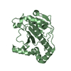
| Structure viewer | Molecule: MolmilJmol/JSmol |
|---|
- Downloads & links
-Download
| PDBx/mmCIF format | 1nrf.cif.gz | 63.9 KB | Display | PDBx/mmCIF format |
|---|---|---|---|---|
| PDB format | pdb1nrf.ent.gz | 47.2 KB | Display | PDB format |
| PDBx/mmJSON format | 1nrf.json.gz | Tree view | PDBx/mmJSON format | |
| Others |  Other downloads Other downloads |
-Validation report
| Arichive directory | https://data.pdbj.org/pub/pdb/validation_reports/nr/1nrfftp://data.pdbj.org/pub/pdb/validation_reports/nr/1nrf | HTTPS FTP |
|---|
-Related structure data
| Similar structure data |
|---|


-Links
 PDBj
PDBj
- Assembly
| Deposited unit | 
| ||||||||
|---|---|---|---|---|---|---|---|---|---|
| 1 |
| ||||||||
| Unit cell |
|
-Components
| #1: Protein | Mass: 29902.072 Da / Num. of mol.: 1 / Fragment: C-terminal domain Source method: isolated from a genetically manipulated source Source: (gene. exp.) |
|---|---|
| #2: Water | ChemComp-HOH /  Mass: 18.015 Da / Num. of mol.: 81 / Source method: isolated from a natural source / Formula: H2O Mass: 18.015 Da / Num. of mol.: 81 / Source method: isolated from a natural source / Formula: H2O |
-Experimental details
-Experiment
| Experiment | Method: X-RAY DIFFRACTION / Number of used crystals: 1 |
|---|
- Sample preparation
| Crystal | Density Matthews: 2.34 Å3/Da / Density % sol: 47.11 % | ||||||||||||||||||||||||||||||||||||||||||
|---|---|---|---|---|---|---|---|---|---|---|---|---|---|---|---|---|---|---|---|---|---|---|---|---|---|---|---|---|---|---|---|---|---|---|---|---|---|---|---|---|---|---|---|
| Crystal grow | Temperature: 293 K / Method: vapor diffusion, hanging drop / pH: 6.95 Details: 18% PEG 8000, 5% Glycerol, 50mM CaCl2 in 0.1M Cacodylate, pH 6.95, VAPOR DIFFUSION, HANGING DROP, temperature 293K | ||||||||||||||||||||||||||||||||||||||||||
| Crystal grow | *PLUS Temperature: 293 K / Method: vapor diffusion, hanging drop | ||||||||||||||||||||||||||||||||||||||||||
| Components of the solutions | *PLUS
|
-Data collection
| Diffraction | Mean temperature: 100 K |
|---|---|
| Diffraction source | Source: SYNCHROTRON / Site: ESRF  / Beamline: BM30A / Wavelength: 0.9786 Å / Beamline: BM30A / Wavelength: 0.9786 Å |
| Detector | Type: MARRESEARCH / Detector: CCD / Date: Sep 7, 2002 |
| Radiation | Protocol: SINGLE WAVELENGTH / Monochromatic (M) / Laue (L): M / Scattering type: x-ray |
| Radiation wavelength | Wavelength: 0.9786 Å / Relative weight: 1 |
| Reflection | Resolution: 2.44→32.444 Å / Num. all: 9979 / Num. obs: 9979 / % possible obs: 99.7 % / Observed criterion σ(F): 0 / Observed criterion σ(I): 0 / Redundancy: 4 % / Biso Wilson estimate: 40.3 Å2 / Rmerge(I) obs: 0.069 / Rsym value: 0.059 / Net I/σ(I): 8.9 |
| Reflection shell | Resolution: 2.44→2.5 Å / Redundancy: 4 % / Rmerge(I) obs: 0.232 / Mean I/σ(I) obs: 3.7 / Num. unique all: 745 / Rsym value: 0.201 / % possible all: 100 |
| Reflection | *PLUS Lowest resolution: 32.44 Å / Rmerge(I) obs: 0.068 |
| Reflection shell | *PLUS % possible obs: 99.9 % / Num. unique obs: 745 |
- Processing
| Software |
| ||||||||||||||||||||||||||||||||||||
|---|---|---|---|---|---|---|---|---|---|---|---|---|---|---|---|---|---|---|---|---|---|---|---|---|---|---|---|---|---|---|---|---|---|---|---|---|---|
| Refinement | Method to determine structure: MOLECULAR REPLACEMENT Starting model: Class D beta-lactamase OXA-2 from Salmonella typhimurium Resolution: 2.5→31.52 Å / Rfactor Rfree error: 0.012 / Isotropic thermal model: RESTRAINED / Cross valid method: THROUGHOUT / σ(F): 2 / Stereochemistry target values: Engh & Huber
| ||||||||||||||||||||||||||||||||||||
| Solvent computation | Solvent model: FLAT MODEL / Bsol: 53.7379 Å2 / ksol: 0.367659 e/Å3 | ||||||||||||||||||||||||||||||||||||
| Displacement parameters | Biso mean: 40 Å2
| ||||||||||||||||||||||||||||||||||||
| Refine analyze |
| ||||||||||||||||||||||||||||||||||||
| Refinement step | Cycle: LAST / Resolution: 2.5→31.52 Å
| ||||||||||||||||||||||||||||||||||||
| Refine LS restraints |
| ||||||||||||||||||||||||||||||||||||
| LS refinement shell | Resolution: 2.5→2.66 Å / Rfactor Rfree error: 0.036 / Total num. of bins used: 6
| ||||||||||||||||||||||||||||||||||||
| Xplor file |
| ||||||||||||||||||||||||||||||||||||
| Refinement | *PLUS Highest resolution: 2.5 Å / Lowest resolution: 31.5 Å / % reflection Rfree: 5.1 % | ||||||||||||||||||||||||||||||||||||
| Solvent computation | *PLUS | ||||||||||||||||||||||||||||||||||||
| Displacement parameters | *PLUS | ||||||||||||||||||||||||||||||||||||
| Refine LS restraints | *PLUS
|
