 Movie
Movie Controller

[English] 日本語
 Yorodumi
- PDB-1nqo: Glyceraldehyde-3-Phosphate Dehydrogenase Mutant With Cys 149 Repl... -
+ Open data

- Basic information
| Entry | Database: PDB / ID: 1nqo | ||||||
|---|---|---|---|---|---|---|---|
| Title | Glyceraldehyde-3-Phosphate Dehydrogenase Mutant With Cys 149 Replaced By Ser Complexed With Nad+ and D-Glyceraldehyde-3-Phosphate | ||||||
 Components Components | Glyceraldehyde 3-phosphate dehydrogenase | ||||||
 Keywords Keywords | OXIDOREDUCTASE / Glycolysis / NAD | ||||||
| Function / homology |  Function and homology information Function and homology informationglyceraldehyde-3-phosphate dehydrogenase (phosphorylating) / glyceraldehyde-3-phosphate dehydrogenase (NAD+) (phosphorylating) activity / glycolytic process / glucose metabolic process / NAD binding / NADP binding / cytoplasm Similarity search - Function | ||||||
| Biological species |   Geobacillus stearothermophilus (bacteria) Geobacillus stearothermophilus (bacteria) | ||||||
| Method |  X-RAY DIFFRACTION / FOURIER SYNTHESIS / Resolution: 2.01 Å X-RAY DIFFRACTION / FOURIER SYNTHESIS / Resolution: 2.01 Å | ||||||
 Authors Authors | Didierjean, C. / Corbier, C. / Fatih, M. / Favier, F. / Boschi-Muller, S. / Branlant, G. / Aubry, A. | ||||||
 Citation Citation | Journal: J.Biol.Chem. / Year: 2003 Title: Crystal structure of two ternary complexes of phosphorylating Glyceraldehyde-3-Phosphate Dehydrogenase from Bacillus stearothermophilus with NAD and D-Glyceraldehyde-3-Phosphate Authors: Didierjean, C. / Corbier, C. / Fatih, M. / Favier, F. / Boschi-Muller, S. / Branlant, G. / Aubry, A. | ||||||
| History |
|
- Structure visualization
| Structure viewer | Molecule: MolmilJmol/JSmol |
|---|
- Downloads & links
-Download
| PDBx/mmCIF format | 1nqo.cif.gz | 276.5 KB | Display | PDBx/mmCIF format |
|---|---|---|---|---|
| PDB format | pdb1nqo.ent.gz | 222.7 KB | Display | PDB format |
| PDBx/mmJSON format | 1nqo.json.gz | Tree view | PDBx/mmJSON format | |
| Others |  Other downloads Other downloads |
-Validation report
| Arichive directory | https://data.pdbj.org/pub/pdb/validation_reports/nq/1nqoftp://data.pdbj.org/pub/pdb/validation_reports/nq/1nqo | HTTPS FTP |
|---|
-Related structure data
| Related structure data | 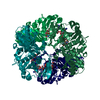 1nptC 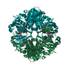 1nq5SC 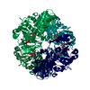 1nqaC C: citing same article ( S: Starting model for refinement |
|---|---|
| Similar structure data |


-Links
 PDBj
PDBj
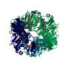
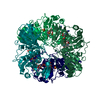
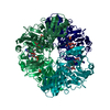
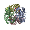
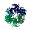
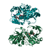
- Assembly
| Deposited unit | 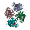
| ||||||||
|---|---|---|---|---|---|---|---|---|---|
| 1 | 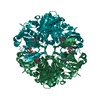
| ||||||||
| 2 | 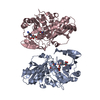
| ||||||||
| 3 | 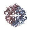
| ||||||||
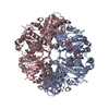
| Unit cell |
| ||||||||
| Details | The second part of the first biological tetramer is generated by the symmetry operator (1-x, y, 2-z). / The second part of the second biological tetramer is generated by the symmetry operator (-x, y, 1-z). |
-Components
| #1: Protein | Mass: 35974.984 Da / Num. of mol.: 4 / Mutation: C149S Source method: isolated from a genetically manipulated source Source: (gene. exp.) Geobacillus stearothermophilus (bacteria)Gene: GAP / Plasmid: pBluescriptII / Production host: References: UniProt: P00362, glyceraldehyde-3-phosphate dehydrogenase (phosphorylating) #2: Chemical | ChemComp-NAD /   Mass: 663.425 Da / Num. of mol.: 4 / Source method: obtained synthetically / Formula: C21H27N7O14P2 / Comment: NAD*YM Mass: 663.425 Da / Num. of mol.: 4 / Source method: obtained synthetically / Formula: C21H27N7O14P2 / Comment: NAD*YM#3: Chemical | ChemComp-G3H / 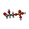  Mass: 170.058 Da / Num. of mol.: 4 / Source method: obtained synthetically / Formula: C3H7O6P Mass: 170.058 Da / Num. of mol.: 4 / Source method: obtained synthetically / Formula: C3H7O6P#4: Water | ChemComp-HOH / |  Mass: 18.015 Da / Num. of mol.: 675 / Source method: isolated from a natural source / Formula: H2O Mass: 18.015 Da / Num. of mol.: 675 / Source method: isolated from a natural source / Formula: H2O |
|---|
-Experimental details
-Experiment
| Experiment | Method: X-RAY DIFFRACTION / Number of used crystals: 1 |
|---|
- Sample preparation
| Crystal | Density Matthews: 2.09 Å3/Da / Density % sol: 40.69 % | ||||||||||||||||||||||||||||||||||||||||||||||||
|---|---|---|---|---|---|---|---|---|---|---|---|---|---|---|---|---|---|---|---|---|---|---|---|---|---|---|---|---|---|---|---|---|---|---|---|---|---|---|---|---|---|---|---|---|---|---|---|---|---|
| Crystal grow | Temperature: 293 K / Method: vapor diffusion, hanging drop / pH: 7.5 Details: PEG 4000, sodium acetate, Tris-HCl, pH 7.5, VAPOR DIFFUSION, HANGING DROP, temperature 293K | ||||||||||||||||||||||||||||||||||||||||||||||||
| Crystal grow | *PLUS pH: 8.9 | ||||||||||||||||||||||||||||||||||||||||||||||||
| Components of the solutions | *PLUS
|
-Data collection
| Diffraction | Mean temperature: 100 K |
|---|---|
| Diffraction source | Source: ROTATING ANODE / Type: ENRAF-NONIUS FR591 / Wavelength: 1.5418 Å |
| Detector | Type: MAC Science DIP-2030B / Detector: IMAGE PLATE / Date: Mar 17, 1999 / Details: MIRRORS |
| Radiation | Monochromator: MIRRORS / Protocol: SINGLE WAVELENGTH / Monochromatic (M) / Laue (L): M / Scattering type: x-ray |
| Radiation wavelength | Wavelength: 1.5418 Å / Relative weight: 1 |
| Reflection | Resolution: 2.01→20 Å / Num. all: 78775 / Num. obs: 78775 / % possible obs: 92.8 % / Observed criterion σ(I): 0 / Rmerge(I) obs: 0.105 / Net I/σ(I): 12.1 |
| Reflection shell | Resolution: 2.01→2.07 Å / Rmerge(I) obs: 0.32 / Mean I/σ(I) obs: 3.2 / % possible all: 74.8 |
| Reflection | *PLUS Num. obs: 77562 |
| Reflection shell | *PLUS % possible obs: 74.8 % |
- Processing
| Software |
| ||||||||||||||||||||||||
|---|---|---|---|---|---|---|---|---|---|---|---|---|---|---|---|---|---|---|---|---|---|---|---|---|---|
| Refinement | Method to determine structure: FOURIER SYNTHESIS Starting model: PDB ENTRY 1NQ5 Resolution: 2.01→8 Å / σ(F): 0 / Stereochemistry target values: Engh & Huber
| ||||||||||||||||||||||||
| Refinement step | Cycle: LAST / Resolution: 2.01→8 Å
| ||||||||||||||||||||||||
| LS refinement shell | Resolution: 2.01→2.07 Å /
| ||||||||||||||||||||||||
| Refinement | *PLUS % reflection Rfree: 10 % / Rfactor Rfree: 0.235 | ||||||||||||||||||||||||
| Solvent computation | *PLUS | ||||||||||||||||||||||||
| Displacement parameters | *PLUS | ||||||||||||||||||||||||
| Refine LS restraints | *PLUS
|
