 Movie
Movie Controller

[English] 日本語
 Yorodumi
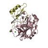
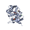
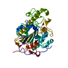
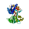
- PDB-1nnt: STRUCTURAL EVIDENCE FOR A PH-SENSITIVE DI-LYSINE TRIGGER IN THE H... -
+ Open data

- Basic information
| Entry | Database: PDB / ID: 1nnt | ||||||
|---|---|---|---|---|---|---|---|
| Title | STRUCTURAL EVIDENCE FOR A PH-SENSITIVE DI-LYSINE TRIGGER IN THE HEN OVOTRANSFERRIN N-LOBE: IMPLICATIONS FOR TRANSFERRIN IRON RELEASE | ||||||
 Components Components | OVOTRANSFERRIN | ||||||
 Keywords Keywords | IRON TRANSPORT PROTEIN | ||||||
| Function / homology |  Function and homology information Function and homology informationorganomineral extracellular matrix / iron ion transmembrane transport / antimicrobial humoral response / ferric iron binding / acute-phase response / iron ion transport / recycling endosome / antibacterial humoral response / response to lipopolysaccharide / intracellular iron ion homeostasis ...organomineral extracellular matrix / iron ion transmembrane transport / antimicrobial humoral response / ferric iron binding / acute-phase response / iron ion transport / recycling endosome / antibacterial humoral response / response to lipopolysaccharide / intracellular iron ion homeostasis / early endosome / iron ion binding / response to xenobiotic stimulus / : / plasma membrane Similarity search - Function | ||||||
| Biological species |  | ||||||
| Method |  X-RAY DIFFRACTION / Resolution: 2.3 Å X-RAY DIFFRACTION / Resolution: 2.3 Å | ||||||
 Authors Authors | Dewan, J.C. / Mikami, B. / Sacchettini, J.C. | ||||||
 Citation Citation | Journal: Biochemistry / Year: 1993 Title: Structural evidence for a pH-sensitive dilysine trigger in the hen ovotransferrin N-lobe: implications for transferrin iron release. Authors: Dewan, J.C. / Mikami, B. / Hirose, M. / Sacchettini, J.C. | ||||||
| History |
| ||||||
| Remark 650 | HELIX HELIX NAMES ARE SIMILAR TO HUMAN LACTOFERRIN (TABLE 3, J. MOL. BIOL., VOL. 209, P. 711, 1993) ...HELIX HELIX NAMES ARE SIMILAR TO HUMAN LACTOFERRIN (TABLE 3, J. MOL. BIOL., VOL. 209, P. 711, 1993) WHICH ARE SIMILAR TO THE RABBIT SERUM TRANSFERRIN STRUCTURE (BIOCHEMISTRY, VOL. 27, P. 5804, 1988). HELIX NAMES FOR RESIDUES 167 - 174 ARE NOT NOTED IN LACTOFERRIN STRUCTURE. | ||||||
| Remark 700 | SHEET IN SHEET RECORDS BELOW, STRAND NAMES ARE SAME AS HUMAN LACTOFERRIN (TABLE 3, J. MOL. BIOL., ...SHEET IN SHEET RECORDS BELOW, STRAND NAMES ARE SAME AS HUMAN LACTOFERRIN (TABLE 3, J. MOL. BIOL., VOL. 209, P. 711, 1993) WHICH ARE SAME AS THE RABBIT SERUM TRANSFERRIN STRUCTURE (BIOCHEMISTRY, VOL. 27, P. 5804, 1988). |
- Structure visualization
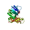
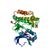
| Structure viewer | Molecule: MolmilJmol/JSmol |
|---|
- Downloads & links
-Download
| PDBx/mmCIF format | 1nnt.cif.gz | 79.4 KB | Display | PDBx/mmCIF format |
|---|---|---|---|---|
| PDB format | pdb1nnt.ent.gz | 58.4 KB | Display | PDB format |
| PDBx/mmJSON format | 1nnt.json.gz | Tree view | PDBx/mmJSON format | |
| Others |  Other downloads Other downloads |
-Validation report
| Arichive directory | https://data.pdbj.org/pub/pdb/validation_reports/nn/1nntftp://data.pdbj.org/pub/pdb/validation_reports/nn/1nnt | HTTPS FTP |
|---|
-Related structure data
| Similar structure data |
|---|


-Links
 PDBj
PDBj


- Assembly
| Deposited unit | 
| ||||||||
|---|---|---|---|---|---|---|---|---|---|
| 1 |
| ||||||||
| Unit cell |
| ||||||||
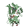
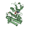
| Atom site foot note | 1: CIS PROLINE - PRO 71 / 2: CIS PROLINE - PRO 287 3: THERE IS A STRONG 2.3 ANGSTROMS H-BOND BETWEEN NZ OF LYS 209 AND NZ OF LYS 301 WHICH IT IS SUGGESTED FORMS A PH-SENSITIVE DI-LYSINE TRIGGER THAT OPENS THE TWO DOMAINS OF THE N-LOBE AT LOW PH AND ...3: THERE IS A STRONG 2.3 ANGSTROMS H-BOND BETWEEN NZ OF LYS 209 AND NZ OF LYS 301 WHICH IT IS SUGGESTED FORMS A PH-SENSITIVE DI-LYSINE TRIGGER THAT OPENS THE TWO DOMAINS OF THE N-LOBE AT LOW PH AND FACILITATES FE(III) RELEASE. |
-Components
| #1: Protein | Mass: 36105.773 Da / Num. of mol.: 1 Source method: isolated from a genetically manipulated source Source: (gene. exp.) |
|---|---|
| #2: Chemical | ChemComp-FE /   Mass: 55.845 Da / Num. of mol.: 1 / Source method: obtained synthetically / Formula: Fe Mass: 55.845 Da / Num. of mol.: 1 / Source method: obtained synthetically / Formula: Fe |
| #3: Chemical | ChemComp-CO3 /   Mass: 60.009 Da / Num. of mol.: 1 / Source method: obtained synthetically / Formula: CO3 Mass: 60.009 Da / Num. of mol.: 1 / Source method: obtained synthetically / Formula: CO3 |
| #4: Water | ChemComp-HOH /  Mass: 18.015 Da / Num. of mol.: 108 / Source method: isolated from a natural source / Formula: H2O Mass: 18.015 Da / Num. of mol.: 108 / Source method: isolated from a natural source / Formula: H2O |
| Compound details | THERE IS A STRONG 2.3 ANGSTROMS H-BOND BETWEEN NZ OF LYS 209 AND NZ OF LYS 301 WHICH IT IS ...THERE IS A STRONG 2.3 ANGSTROMS H-BOND BETWEEN NZ OF LYS 209 AND NZ OF LYS 301 WHICH IT IS SUGGESTED FORMS A PH-SENSITIVE DI-LYSINE TRIGGER THAT OPENS THE TWO DOMAINS OF THE N-LOBE AT LOW PH AND FACILITATE |
| Has protein modification | Y |
| Sequence details | SEQUENCE ADVISORY NOTICE DIFFERENCE BETWEEN SWISS-PROT AND PDB SEQUENCE. SWISS-PROT ENTRY NAME: ...SEQUENCE ADVISORY NOTICE DIFFERENCE |
-Experimental details
-Experiment
| Experiment | Method: X-RAY DIFFRACTION |
|---|
- Sample preparation
| Crystal | Density Matthews: 2.26 Å3/Da / Density % sol: 45.6 % | ||||||||||||||||||||||||||||||||||||||||||||||||
|---|---|---|---|---|---|---|---|---|---|---|---|---|---|---|---|---|---|---|---|---|---|---|---|---|---|---|---|---|---|---|---|---|---|---|---|---|---|---|---|---|---|---|---|---|---|---|---|---|---|
| Crystal grow | *PLUS pH: 5.9 / Method: vapor diffusion, hanging drop / Details: used to seed | ||||||||||||||||||||||||||||||||||||||||||||||||
| Components of the solutions | *PLUS
|
-Data collection
| Radiation | Scattering type: x-ray |
|---|---|
| Radiation wavelength | Relative weight: 1 |
| Reflection | *PLUS Highest resolution: 2.3 Å / Num. obs: 14266 / % possible obs: 94 % / Observed criterion σ(I): 8.6 / Num. measured all: 59108 / Rmerge(I) obs: 0.09 |
- Processing
| Software |
| ||||||||||||||||||||||||||||||||||||||||||||||||||||||||||||
|---|---|---|---|---|---|---|---|---|---|---|---|---|---|---|---|---|---|---|---|---|---|---|---|---|---|---|---|---|---|---|---|---|---|---|---|---|---|---|---|---|---|---|---|---|---|---|---|---|---|---|---|---|---|---|---|---|---|---|---|---|---|
| Refinement | Rfactor Rwork: 0.16 / Rfactor obs: 0.16 / Highest resolution: 2.3 Å | ||||||||||||||||||||||||||||||||||||||||||||||||||||||||||||
| Refinement step | Cycle: LAST / Highest resolution: 2.3 Å
| ||||||||||||||||||||||||||||||||||||||||||||||||||||||||||||
| Refine LS restraints |
| ||||||||||||||||||||||||||||||||||||||||||||||||||||||||||||
| Refinement | *PLUS Lowest resolution: 20 Å / Num. reflection obs: 8965 / σ(I): 2 / Rfactor obs: 0.16 / Rfactor Rwork: 0.16 | ||||||||||||||||||||||||||||||||||||||||||||||||||||||||||||
| Solvent computation | *PLUS | ||||||||||||||||||||||||||||||||||||||||||||||||||||||||||||
| Displacement parameters | *PLUS | ||||||||||||||||||||||||||||||||||||||||||||||||||||||||||||
| Refine LS restraints | *PLUS Type: x_angle_d / Dev ideal: 2.25 |
