 Movie
Movie Controller

[English] 日本語
 Yorodumi
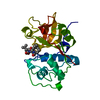
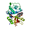
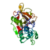
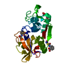
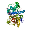
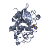
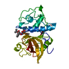
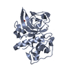
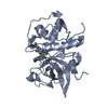
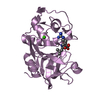
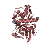
- PDB-1nl6: Crystal Structure Of The Cysteine Protease Human Cathepsin K In C... -
+ Open data

- Basic information
| Entry | Database: PDB / ID: 1nl6 | ||||||
|---|---|---|---|---|---|---|---|
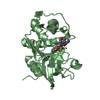
| Title | Crystal Structure Of The Cysteine Protease Human Cathepsin K In Complex With A Covalent Azepanone Inhibitor | ||||||
 Components Components | Cathepsin K | ||||||
 Keywords Keywords | HYDROLASE / SULFHYDRYL PROTEINASE | ||||||
| Function / homology |  Function and homology information Function and homology informationcathepsin K / negative regulation of cartilage development / RUNX1 regulates transcription of genes involved in differentiation of keratinocytes / endolysosome lumen / thyroid hormone generation / Trafficking and processing of endosomal TLR / proteoglycan binding / Activation of Matrix Metalloproteinases / Collagen degradation / collagen catabolic process ...cathepsin K / negative regulation of cartilage development / RUNX1 regulates transcription of genes involved in differentiation of keratinocytes / endolysosome lumen / thyroid hormone generation / Trafficking and processing of endosomal TLR / proteoglycan binding / Activation of Matrix Metalloproteinases / Collagen degradation / collagen catabolic process / fibronectin binding / extracellular matrix disassembly / bone resorption / mitophagy / collagen binding / Degradation of the extracellular matrix / cysteine-type peptidase activity / MHC class II antigen presentation / lysosomal lumen / : / lysosome / apical plasma membrane / serine-type endopeptidase activity / external side of plasma membrane / cysteine-type endopeptidase activity / proteolysis / : / extracellular region / nucleoplasm Similarity search - Function | ||||||
| Biological species |  Homo sapiens (human) Homo sapiens (human) | ||||||
| Method |  X-RAY DIFFRACTION / SYNCHROTRON / MOLECULAR REPLACEMENT / Resolution: 2.8 Å X-RAY DIFFRACTION / SYNCHROTRON / MOLECULAR REPLACEMENT / Resolution: 2.8 Å | ||||||
 Authors Authors | Smith, W.W. / Janson, C.A. / Zhao, B. | ||||||
 Citation Citation | Journal: J.Med.Chem. / Year: 2001 Title: Azepanone-based inhibitors of human and rat cathepsin K Authors: Marquis, R.W. / Ru, Y. / LoCastro, S.M. / Zeng, J. / Yamashita, D.S. / Oh, H.J. / Erhard, K.F. / Davis, L.D. / Tomaszek, T.A. / Tew, D. / Salyers, K. / Proksch, J. / Ward, K. / Smith, B. / ...Authors: Marquis, R.W. / Ru, Y. / LoCastro, S.M. / Zeng, J. / Yamashita, D.S. / Oh, H.J. / Erhard, K.F. / Davis, L.D. / Tomaszek, T.A. / Tew, D. / Salyers, K. / Proksch, J. / Ward, K. / Smith, B. / Levy, M. / Cummings, M.D. / Haltiwanger, R.C. / Trescher, G. / Wang, B. / Hemling, M.E. / Quinn, C.J. / Cheng, H.-Y. / Lin, F. / Smith, W.W. / Janson, C.A. / Zhao, B. / McQueney, M.S. / D'Alessio, K. / Lee, C.P. / Marzulli, A. / A Dodds, R. / Blake, S. / Hwang, S.M. / James, I.E. / Gress, C.J. / Bradley, B.R. / Lark, M.W. / Gowen, M. / Veber, D.F. | ||||||
| History |
|
- Structure visualization
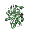
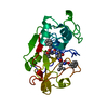
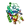
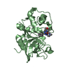
| Structure viewer | Molecule: MolmilJmol/JSmol |
|---|
- Downloads & links
-Download
| PDBx/mmCIF format | 1nl6.cif.gz | 95.8 KB | Display | PDBx/mmCIF format |
|---|---|---|---|---|
| PDB format | pdb1nl6.ent.gz | 74.4 KB | Display | PDB format |
| PDBx/mmJSON format | 1nl6.json.gz | Tree view | PDBx/mmJSON format | |
| Others |  Other downloads Other downloads |
-Validation report
| Arichive directory | https://data.pdbj.org/pub/pdb/validation_reports/nl/1nl6ftp://data.pdbj.org/pub/pdb/validation_reports/nl/1nl6 | HTTPS FTP |
|---|
-Related structure data
| Related structure data |  1nljC 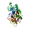 1atkS S: Starting model for refinement C: citing same article ( |
|---|---|
| Similar structure data |


-Links
 PDBj
PDBj


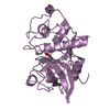
- Assembly
| Deposited unit | 
| ||||||||
|---|---|---|---|---|---|---|---|---|---|
| 1 | 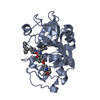
| ||||||||
| 2 | 
| ||||||||
| Unit cell |
|
-Components
| #1: Protein | Mass: 23523.480 Da / Num. of mol.: 2 Source method: isolated from a genetically manipulated source Source: (gene. exp.) Homo sapiens (human) / Cell line (production host): SF9 / Production host:   Spodoptera frugiperda (fall armyworm) / References: UniProt: P43235, cathepsin K Spodoptera frugiperda (fall armyworm) / References: UniProt: P43235, cathepsin K#2: Chemical |   Mass: 709.830 Da / Num. of mol.: 2 / Source method: obtained synthetically / Formula: C40H47N5O7 Mass: 709.830 Da / Num. of mol.: 2 / Source method: obtained synthetically / Formula: C40H47N5O7Has protein modification | Y | |
|---|
-Experimental details
-Experiment
| Experiment | Method: X-RAY DIFFRACTION / Number of used crystals: 1 |
|---|
- Sample preparation
| Crystal | Density Matthews: 3.32 Å3/Da / Density % sol: 63 % | ||||||||||||||||||||||||
|---|---|---|---|---|---|---|---|---|---|---|---|---|---|---|---|---|---|---|---|---|---|---|---|---|---|
| Crystal grow | Temperature: 298 K / Method: vapor diffusion, sitting drop / pH: 8 Details: 10% PEG 8000, 0.1 M imidazole, 0.2 M calcium acetate, pH 8.0, VAPOR DIFFUSION, SITTING DROP, temperature 298K | ||||||||||||||||||||||||
| Crystal grow | *PLUS Method: vapor diffusion | ||||||||||||||||||||||||
| Components of the solutions | *PLUS
|
-Data collection
| Diffraction | Mean temperature: 100 K |
|---|---|
| Diffraction source | Source: SYNCHROTRON / Site: APS  / Beamline: 17-ID / Wavelength: 1 Å / Beamline: 17-ID / Wavelength: 1 Å |
| Detector | Type: MARRESEARCH / Detector: CCD / Date: Mar 1, 2000 |
| Radiation | Monochromator: Si 111 / Protocol: SINGLE WAVELENGTH / Monochromatic (M) / Laue (L): M / Scattering type: x-ray |
| Radiation wavelength | Wavelength: 1 Å / Relative weight: 1 |
| Reflection | Resolution: 2.55→99 Å / Num. all: 19740 / Num. obs: 17372 / % possible obs: 88 % / Observed criterion σ(F): 2 / Observed criterion σ(I): 2 / Redundancy: 9.8 % / Rmerge(I) obs: 0.58 / Rsym value: 0.58 / Net I/σ(I): 32 |
| Reflection shell | Resolution: 2.55→2.59 Å / Rmerge(I) obs: 0.94 / Mean I/σ(I) obs: 9.9 / Num. unique all: 631 / Rsym value: 0.94 / % possible all: 65 |
| Reflection | *PLUS Lowest resolution: 99 Å / % possible obs: 88 % |
- Processing
| Software |
| |||||||||||||||||||||||||
|---|---|---|---|---|---|---|---|---|---|---|---|---|---|---|---|---|---|---|---|---|---|---|---|---|---|---|
| Refinement | Method to determine structure: MOLECULAR REPLACEMENT Starting model: PDB ENTRY 1ATK Resolution: 2.8→6 Å / Cross valid method: THROUGHOUT / σ(F): 2 / Stereochemistry target values: Engh & Huber
| |||||||||||||||||||||||||
| Displacement parameters |
| |||||||||||||||||||||||||
| Refinement step | Cycle: LAST / Resolution: 2.8→6 Å
| |||||||||||||||||||||||||
| Refine LS restraints |
| |||||||||||||||||||||||||
| LS refinement shell | Resolution: 2.8→2.83 Å
| |||||||||||||||||||||||||
| Xplor file |
| |||||||||||||||||||||||||
| Refinement | *PLUS Lowest resolution: 6 Å / % reflection Rfree: 10 % / Rfactor Rwork: 0.26 | |||||||||||||||||||||||||
| Solvent computation | *PLUS | |||||||||||||||||||||||||
| Displacement parameters | *PLUS | |||||||||||||||||||||||||
| Refine LS restraints | *PLUS Type: c_bond_d / Dev ideal: 0.01 |
